Optical trap 'grabs and scans' bacterium
Scientists from the Department of Microsystems Engineering (IMTEK) of the University of Freiburg (Freiburg, Germany) have developed a laser-driven optical trap that can grab and scan tiny, elongated bacteria.
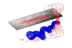
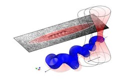
Physicists Prof. Dr. Alexander Rohrbach and Matthias Koch created a kind of light tube that traps the agile unicellular organisms. Optical tweezers could previously only be used to grab bacteria at one point, not to manipulate their orientation. The researchers have now succeeded in using a quickly moving, focused laser beam to exert an equally distributed force over the entire bacterium, which constantly changes its complex form. At the same time, they were able to record the movements of the trapped bacterium in high-speed, three-dimensional (3D) images by measuring miniscule deflections of the light particles.
The scientists investigated so-called spiroplasmas in their study. These spiral-shaped bacteria have a diameter of only 200 nm, roughly equivalent to the thickness of 1,000 atoms. Since they do not have a cell wall, they can rapidly change their shape and thereby move. It is not possible to image these bacteria sufficiently through a conventional optical microscope due to their small size and their rapid movements. Using the newly developed light-scanning optical trap, however, the biophysicists were able to hold and orient the bacterium over its entire length.
One important attribute of laser light is that interfering light particles can increase or decrease their brightness. When light hits the bacterium and is deflected from it, this light superposes with the non-deflected light and thus is amplified, enabling the generation of three-dimensional images that do not only have a high contrast but also an increased resolution. In this way, it is possible to acquire up to 1,000 3D images/s and record the rapid movements of the bacterium in detail in a movie.
In the future, the scientists plan to use this method to study the behavior and cellular mechanics of other bacteria that do not have a cell wall and that are therefore difficult to treat with antibiotics. These studies could thus help scientists to reach a better understanding of bacterial infectious diseases.
The team's findings have been published in Nature Photonics; for more information, please visit http://www.nature.com/doifinder/10.1038/nphoton.2012.232.
-----
Follow us on Twitter, 'like' us on Facebook, and join our group on LinkedIn
Laser Focus World has gone mobile: Get all of the mobile-friendly options here.
Subscribe now to BioOptics World magazine; it's free!

