Nanosecond lasers diagnose melanoma without tears
A team at the optics firm Lasertechnik Berlin (LTB; Berlin, Germany) has developed a laser-based method of identifying the disease. So far the team has applied the approach only to tissue samples. But it should soon be possible to apply the method directly to patients in situ, without removing any skin.
The ability to diagnose malignant melanoma without physical biopsies would negate an issue that concerns the medical profession. Because the disease has a high fatality rate—accounting for about 48,000 deaths each year worldwide—dermatologists are quick to sample patients’ moles and other suspect skin areas for biopsy. However, studies have shown that 90% of all those biopsies prove negative for malignant melanoma. A nondestructive diagnostic method would save numerous patients unnecessary pain and inconvenience.
Melanoma is cancer of the melanocytes. A few of these pigment-producing cells inhabit the bowels and the eyes, but they exist mainly in the skin. There, the melanocytes produce melanin, a pigment that fluoresces. Inducing such fluorescence by a femtosecond laser would theoretically allow scientists to detect changes in the melanin that indicate the presence of cancer. Unfortunately, fluorescence from other skin components largely masks that from melanin.
Two years ago, LTB found a way around that problem. “We lengthened the laser pulse,” LTB’s managing director Matthias Scholz explains. “Nanosecond pulses suppress the disruptive fluorescence to a great extent.”
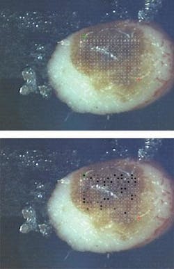
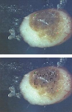
Scientists from LTB and the Dermatology Clinic of the Ruhr University Bochum have applied those pulses to biopsied tissue samples (see figure). “We use a dye laser pumped by a nitrogen laser,” says LTB senior scientist Dieter Leupold. “We measure the melanin-dominated fluorescence of the biopsies immediately after excision in up to 16 spectral channels distributed over the visible range. From the spectral shape we can differentiate whether the pigmented tissue are is benign, malign, or beginning degeneration.”
The group compared its laser-based diagnoses with those derived from histological study of 200 biopsied tissues. “In all cases we had agreement on the diagnosis of malignant melanoma,” Scholz recalls. LTB’s method also revealed small tissue areas with malignant degeneration that histology did not detect.
The technique has also proved successful in diagnosing melanoma of the eye and the less threatening form of skin cancer called pigmented basal cell carcinoma. —PG

