BioOptics BreakthRoughs
Green fiber laser passes NIH tests
Following testing in flow-cytometry applications, the National Cancer Institute gave a green light to Zecotek Photonics’ (Vancouver, B.C.) green fiber laser GFL-550, saying it meets all performance specifications by emitting a 550 nm wavelength.
“Until now a 550 nm solution did not exist,” said Dr. William Telford, Head of the Flow Cytometry Core Laboratory at the National Cancer Institute/NIH, who presented the GFL-550’s performance in a paper by at the 24th International Congress of the International Society for Analytical Cytometry (Budapest, Hungary; May 2008).
Because it excites certain fluorescent probes more efficiently, the new laser promises improved accuracy and broader use of flow cytometry in immunology, hematology, transplants, and biomedical research. Zecotek sees the offering as the replacement of choice for 532 nm lasers in confocal microscopy as well as cytometry. Contact: John Moore, [email protected].
High resolution in real time
The first commercial version of the highest resolution wide-field light microscope is now in place at the University of California–Davis Center for Biophotonics Science and Technology (Sacramento, CA).
Called OMX (short for Optical Microscopy eXperimental), the system allows cellular processes to be viewed at the smallest possible levels—as they occur. It uses a new imaging technology, structured illumination, invented by Mats Gustafsson, a UC San Francisco postdoctoral researcher.
Structured illumination overcomes limitations such as diffraction limit or the inability to provide high-resolution imaging or living samples. With it, a carefully designed pattern of light is used to illuminate an object. The pattern, which looks more like a bar code than a uniform light field, produces moiré patterns that are then resolved by the microscope. Software then digitally reconstructs three-dimensional, ultra-high-resolution images on a computer screen from the multiple individual images generated with the illumination pattern.
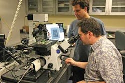
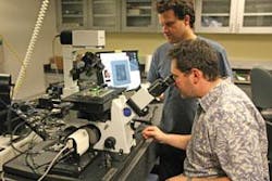
The system actually consists of two microscopes: a DeltaVision unit, which is commercially available from Applied Precision, gives scientists a wide view of the slide and identification of different regions of interest (see photo) that can then be rastered for transfer to the superresolution OMX. The OMX doesn’t have optics to look through, so it is kept inside a cabinet: a computer takes all the images and recreates them on a computer screen. In addition to ultra-high-resolution images, the OMX can produce rapid multiwavelength, three-dimensional images of live samples for the real-time study of cellular processes in action.
The breast cancer research team at UC Davis plans to use OMX to closely evaluate cell changes that support the growth of malignancies. Contact: Dennis Matthews, [email protected].
Novel glaucoma therapy aces clinical study
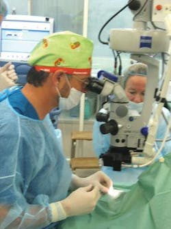
IOPtima’s (Ramat Gan, Israel) laser-based OT134 device and procedure has successfully completed the three-month follow-up period for an initial human clinical study. The technology allows eye surgeons to reduce internal eye pressure without penetrating the eye membrane.
The study involved 13 patients treated at a leading glaucoma center in Mexico City under the supervision of Professor Felix Gil Carrasco. IOPtima intends to further monitor the patients for at least one year.
The study is the first part of a multinational clinical trial at ophthalmology centers around the world aimed at obtaining regulatory approval for Europe. In addition, IOPtima plans to meet with the FDA to initiate procedures aimed at U.S. approval. IOPtima’s aim is to achieve the effect of non-penetrating deep sclerectomy (NPDS) surgery while removing the risk of perforating the membrane and minimizing the risk of perforating the scleral tissue via its CO2 laser-based system. The OT134 is self-terminating once the desired scleral thickness has been achieved: the laser stops ablating as soon as it comes in contact with the intraocular percolated liquid, which occurs as soon as the laser reaches the optimal residual intact layer thickness.
Previously the most efficient surgical approach was trabeculectomy; NPDS is a similar but modified procedure causing a significantly smaller number of side effects. The former involves penetrating through the wall of the eye; doing so without inadvertently perforating the thin trabecular membrane is prohibitive with traditional technologies. Contact: Joshua Degani, [email protected]
Label-free high-throughput milestone
Scientists from two of the top 10 pharmaceutical companies have each screened 100,000 drug candidates using Corning’s Epic high-throughput label-free screening system. These screens exemplify the progress label-free technology has made in the recent years.
Removing fluorescent, luminescent, and radioactive labels from assays promises to improve the speed and quality of the drug discovery process, open a wider variety of drug targets, and enable researchers to study drug candidates in more physiologically relevant systems.
Data from both screens were presented at the Society for Biomolecular Sciences’ Label Free Technologies in Drug Discovery Symposium held in Dresden, Germany, in June. Corning’s Epic system, launched in 2006, claims to be the first high-throughput label-free screening system. It uses patented optical biosensor technology to enable the study of a broad range of biochemical and cell-based targets. Removing fluorescent, luminescent and radioactive labels from assays promises to improve the speed and quality of the drug discovery process. “These screens exemplify the remarkable progress label-free technology has made in the few years and the significant impact it can have in the future,” said Ron Verkleeren, Epic business director. Contact: Evi Kostenis,[email protected]; Martin Coldwell, [email protected].


