Ross Eye Institute presents D-EYE portable ophthalmoscope at AAPOS 2016 conference
Two physicians from the Department of Ophthalmology at the State University of New York at Buffalo, headquartered at the Ross Eye Institute (also in Buffalo), incorporated the D-EYE (Padova, Italy) portable ophthalmoscope system into a recent study to determine if the system was a beneficial alternative for fundus imaging in the pediatric population. The results of the study were presented to the American Association of Pediatric Ophthalmology and Strabismus (AAPOS) at the organization’s annual conference held April 6-10, 2016, in Vancouver, BC, Canada.
Related: Portable ophthalmoscope to get telemedicine, cloud storage, and analytical capabilities
During the course of the study, D-EYE was used with an iPhone 6 to obtain retinal images of pediatric patients in both outpatient and inpatient settings, on dilated and undilated eyes. The physicians found that D-EYE was especially helpful in aiding exams in remote settings and with immobile patients because of its compact and lightweight design, which attaches to iOS and Android devices via a specially designed, lightweight bumper. The device uses the phone's camera lens and an LED light source, which is off-axis from the camera aperture, to illuminate the interior of the eye for examination, resulting in no corneal glare.
D-EYE uses the principle of direct ophthalmology. When the pupil is dilated, the device captures a field of view of approximately 20 degrees in a single fundus image at a distance of 1 cm from the patient's eye. When examining an undilated eye, the field of view is approximately 5-8 degrees. Videos are the preferred method of acquisition, as they allow the user to angle the lens to see more structure of the retina. Users pan the retina, starting from the posterior pole, and then move to the upper, nasal, inferior, and nasal peripheral retina to the equator. Color digital images and videos of the retina can be obtained, encompassing the posterior pole, including the macula, optic disc, and (limited by the field of view) towards the peripheral retina.
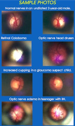
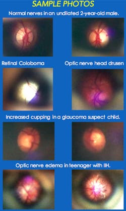
The study concluded that a portable system like D-EYE has a "niche in situations when and where conventional fundus photography is not an option," the physicians involved in the work say. Matthew S. Pihlblad, MD, and Steven G. Stockslager, MD, concluded that the D-EYE system can "document a diffuse range of pathology," including optic nerve, hypoplasia/cupping/pallor, colobomas, optic nerve pits, drusen, and neuroretinitis, among other conditions. The physicians also note the system's clinical utility for general practitioners, pediatricians, emergency physicians, and neurologists, among others, because of the significant potential for telemedicine consultation.
For more information, please visit www.d-eyecare.com.

