3D test promotes early skin cancer detection
Scientists from the Bristol Institute of Technology, University of the West of England, Bristol have developed a 3D test for malignant melanoma that can identify problems not easily spotted in a standard, 2D view of the pattern on the skin.
A group of 2D skin measurements known as the asymmetry, border, color and diameter (ABCD) rules are commonly used to distinguish between malignancy and benign skin growths, but diagnostic accuracy is not convincing.
Now, Lyndon Smith and colleagues at the Machine Vision Laboratory at the Bristol Institute of Technology have collaborated with Robert Warr of the Department of Plastic Surgery, at North Bristol NHS Trust, to develop a computer-assisted diagnosis system that could improve outcomes significantly. The system could quickly and automatically reveal changes in the 3D surface texture of skin that occur in malignant melanoma.
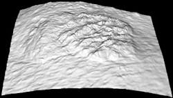
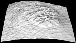
A 3D scan of a skin lesion shows random appearance in the lesion when compared to the more regular surrounding tissue. (Image courtesy Dr. Jiuai Sun, research fellow, Machine Vision Laboratory at the Bristol Institute of Technology)
The team used a handheld, six-light stereo device connected to a laptop computer to scan the skin's surface. This device produces a 3D model of the skin texture patterns. The information is then analyzed by the laptop, which compares it with patterns recorded from known cases of melanoma, used to "train" the software.
Preliminary studies on a sample set including 12 malignant melanomas and 34 benign lesions have given 91.7% sensitivity and 76.4% specificity from analysis of the 3D skin surface normals, the team says.
- "A computer assisted diagnosis system for malignant melanoma using 3D skin surface texture features and artificial neural network," Int. J. Modelling, Identification and Control, 2010, 9, 370-381.
More Brand Name Current Issue Articles
More Brand Name Archives Issue Articles

