OPHTHALMOLOGY/ADAPTIVE OPTICS: Adaptive optics shows earliest evidence of diabetic retinopathy
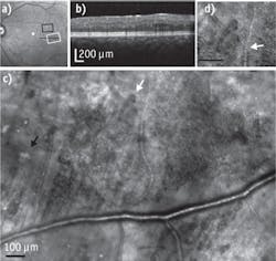
A device able to detect the earliest signs yet of diabetic retinopathy leverages adaptive optics and promises important implications for the millions of Americans dealing with the threat of sight loss related to diabetes.1 Developed at Indiana University (IU; Bloomington, IN) and designed by professor Stephen Burns, associate dean in the School of Optometry, the device uses micro mirrors with moveable segments to reflect light into the eye and to project moving images of the eye's tiny capillaries onto a large screen. Video allows observation of blood cells moving through vessels, while highly magnified retinal images are pieced together with software to create still images.
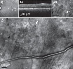
"We had not expected to see such striking changes to the retinas at such early stages," says Ann Elsner, a professor and associate dean in the IU School of Optometry and co-author of the study. "We set out to study the early signs in volunteer research subjects whose eyes were not thought to have very advanced disease. There was damage spread widely across the retina, including changes to blood vessels that were not thought to occur until the more advanced disease states."
Because such changes had not been observable in prior studies, it is not known whether improved control of blood sugar or a change in medications might stop or even reverse the damage. Further research can help determine who has the most severe damage and whether the changes can be reversed.
1. S. A. Burns, Biomed. Opt. Exp., 5, 3, 961 (2014).