Spending even a short time counting things under a microscope tends to inspire dreams of automation. Besides relieving shoulder tension and eye strain, automation of imaging tasks–especially analysis–promises other benefits such as an increase in the number of images processed. Moreover, automated analysis could extract more information from each image–and even compare data across sets of images. A number of academic groups and commercial suppliers are continuing to simplify image analysis.
To push biological imaging ahead, Anne E. Carpenter (see Fig. 1) turned to automation early in her career. As a doctoral student in cell biology at the University of Illinois, Urbana-Champaign, she turned a motorized microscope into an automatic image collector and analyzer. “It became clear that I could count and measure cells for years on end,” she says, “or I could write software to do that for me.” That sort of thinking led Carpenter to her current position as the director of the imaging platform at the Broad Institute (Cambridge, MA), where she and Thouis Jones develop new software tools for imaging.
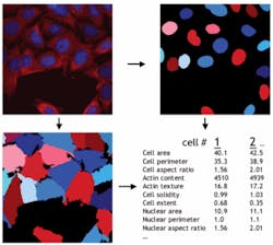
One of those tools, CellProfiler, is an open-source image-analysis application (see Fig. 2). “This is primarily for high-throughput image analysis,” Carpenter explains. It can identify thousands of cells in an image and count or measure them. Moreover, Carpenter and her colleagues developed this piece of software for biologists with no background in computer science. A piece of companion software, CellProfiler Analyst, includes machine-learning capabilities that can even train the main program to count subtypes of cells or cells with particular morphologies.Beyond patterns
Some other programs also use machine learning to get more from images, because part of the challenge in biological imaging comes from the complexity of the samples. “Tissue is very heterogeneous,” says Peter Duncan, director of marketing and business development for life sciences at Definiens (Parsippany, NJ). Consequently, software can’t just look for a pattern. “Generally speaking, biology is all about non-patterns,” Duncan says. “If biology was patterns, we’d have lots of diseases solved.”
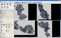
So instead of looking for patterns, the new Definiens Tissue Studio software looks for what Duncan calls associations (see Fig 3). “This software clusters together pixels with similar properties,” Duncan explains. For example, a user can mark an object–say, a lymph node–with a paintbrush tool and then hit a button that causes the program to learn that object. In fact, Definiens Tissue Studio looks at the pixels that make up the object plus those around it. So the software learns to recognize an object in association with things that usually show up nearby. “If I held up a plate in a vacuum, you would call it a white circle,” Duncan says, “but if it’s on a table with a tablecloth and silverware, you would call it a plate, because it is associated with things around it.” Definiens Tissue Studio uses similar associations–but on a tissue scale–to automatically identify objects in a sample.Definiens Tissue Studio can also search for specific dyes or stains. The current version looks for immunohistochemical stains; the next generation will also find immunofluorescence. Duncan describes this software as a tool for translational biomarker research, or moving a biomarker toward pharmaceutical or diagnostic applications. As he says, “It’s for when you really have a hunch that a biomarker is involved with a disease and you want to use that to select patients who will respond to a drug, and measure the marker in a quantitative way.”
Simple sophistication
In bringing such advanced automation to biologists, developers must also consider complexity. “One of today’s biggest challenges is the learning curve for software,” says Kathy Hrach, the product manager at Media Cybernetics (Bethesda, MD). “Most researchers do not have the time to dig in and learn the nuances of a program,” she explains. As a result, Media Cybernetics creates an increasing number of tutorials and educational webinars.
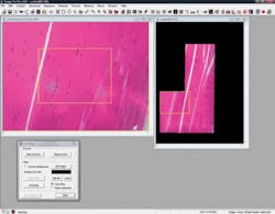
In addition, Media Cybernetics aims at making more-robust products. For example, the recent Image-Pro Plus version 7.0 provides enhanced analytical tools (see Fig. 4). “In general,” says Hrach, “Image-Pro provides the ability to automate manual processes.” For instance, this software can count, classify, and size objects. “It can also track movement of live cells or other images through a frame,” she notes. “The software will even track how far the objects move, or correlate that movement with the movement of other objects.” Beyond improved analytical capabilities, this update brings other improvements. “It includes accelerated control of an x-y-z stage and various modalities,” Hrach says. “It also provides what we call ‘Just in Time’ image loading, which opens large sets of image sequences as fast as opening a single frame.”In the life sciences, however, imaging goes beyond live cells and tissues. In fact, imaging also captures results, especially from molecular biology techniques, including DNA and protein gels, Western blots, and so on. And that’s exactly the area AlphaView Q from Alpha Innotech (San Leandro, CA) targets. Like other advances in biological imaging, though, AlphaView Q aims at simplifying the technology. “It includes an acquisition wizard,” says Lisa Isailovic, marketing manager at Alpha Innotech. “The software asks what you are trying to take a picture of. Is it a gel? Is it a blot?” The wizard also lets a user specify the dye or stain used in an assay. Then, AlphaView Q provides a range of analytical results, including molecular-weight determination, densitometry, and others.
In addition, AlphaView Q helps researchers advance traditional techniques. For example, in the once black-and-white world of Western blots, which identify proteins, researchers can now incorporate various colors of fluorescent dyes, with specific dyes attached to particular proteins. AlphaView Q works with up to three colors at once, and it can even distinguish protein bands of different colors that overlap.
Over the next few years, imaging software will continue to follow this trend of balancing advanced capabilities with user-friendly features. The general idea is to bring powerful imaging abilities to almost anyone who can put this technology to use.
About the Author
Mike May
Contributing Editor, BioOptics World
Mike May writes about instrumentation design and application for BioOptics World. He earned his Ph.D. in neurobiology and behavior from Cornell University and is a member of Sigma Xi: The Scientific Research Society. He has written two books and scores of articles in the field of biomedicine.


