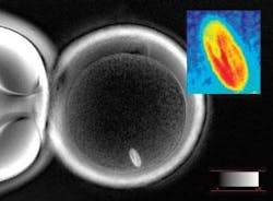
Polarized-light microscope facilitates major stem-cell advance
A microscope with a half century’s worth of improvements played a key role in a major turning point in stem-cell technology: the derivation of embryonic stem cells from reprogrammed monkey skin cells. Scientists at the Oregon National Primate Research Center (Beaverton, OR), who reported the amazing advance last November, used the Oosight noninvasive polarized light microscope from Cambridge Research and Instrumentation (CRi; Woburn, MA) to identify and remove the meiotic spindle and associated genetic material from 304 rhesus monkey eggs. The group then inserted genetic material from the skin cells of an adult male rhesus monkey into the enucleated eggs.
The reprogrammed eggs grew to the blastocyst stage, from which the team produced two stem-cell lines genetically identical to the male monkey. The advance, the first derivation of such stem cells from a primate, points the way to the production of custom tissues that could combat a range of human diseases.
How did the Oosight microscope help? “Before, the problem was always that you could not see the spindle in the egg,” explains team leader Shoukhrat Mitalipov. “The only way to see it was to stain it with dyes. That, we found, was very detrimental for egg quality.” As a further advantage, he adds, “you can actually look in the Oosight microscope and see the spindle with your eyes, not as a frozen computer screen image.”
Technology invented at the Marine Biological Laboratory (MBL; Woods Hole, MA) was used to develop the CRi microscope. MBL cytologist Shinya Inoué pioneered the technology in 1957 when he added a polarization rectifier to a custom-built microscope to improve the contrast and reduce the distortion of images of cells about to undergo division. In the 1990s another MBL group—Rudolf Oldenbourg, Guang Mei, and Michael Shribak—added some next-generation components.
“Liquid crystals are used to switch the polarization of light very quickly to specific states,” Oldenbourg says. “Another key component, an electronic camera, can take images of an object when it is illuminated by any of five different forms of polarized light. And we have developed algorithms that generate an image on your screen with very high contrast that helps to identify spindles.”
Oldenbourg and MBL gynecological researcher David Keefe, now at the University of South Florida (St. Petersburg, FL), then further tweaked the microscope, resulting in a system that reveals plainly visible layers of birefringence around specific targets, such as eggs that researchers want to enucleate (see figure). That set the stage for Mitalipov and his colleagues.
“The Oosight, plus very skilled micromanipulation of the eggs, gave us a 100% success rate with enucleating,” Mitalipov says. —PG
