Two studies validate effectiveness of image calibration software for microscopy

Two recent independent, user-based studies demonstrate that an image calibration software program for microscopy can lead to significant improvements in image quality and consistency for analysis and publication of histological images.
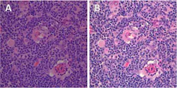
The two papers are important because they provide the first quantitative evidence of both scientific and workflow benefits of a novel colorimetric approach (Datacolor ChromaCal) that provides quality and color consistency across microscope imaging systems and displays. Until recently, reports of the benefits for workflow efficiency, image consistency, and improved analysis had been mostly anecdotal. Pathology and microscopy research imaging have integrated digitization into the workflow, but standards for quality and color consistency from image to image have not yet been set. So many users are looking to research for guidance on how to incorporate these new technologies into their own practice.
Michael A. Linden, MD, Ph.D. (University of Minnesota), and his collaborators reported their findings in the Journal of Histochemistry and Cytochemistry, saying that imaging systems inherently produce image-to-image variations, even if operated by an expert. As a result, post-processing of brightfield images was unavoidable. They studied the performance of both ChromaCal and Adobe Photoshop for delivering image consistency as a precursor to morphometric analysis and concluded that while both systems were effective, Chromacal boosted post-processing in color brightfield imaging to provide a better basis for quantification. It also could be incorporated more readily into workflows, they found.
In another independent study, Dawn M. Dawson, MD, an assistant professor at Case Western Reserve University’s Institute of Pathology, evaluated ChromaCal and reported imaging time savings of more than 90%. Dawson found that image post-processing is necessary to improve histological image comparability for both analysis and manuscript preparation, and said that ChromaCal's objective, colorimetric method avoided tedious and subjective adjustments that typically were needed with traditional image-processing methods. Her case study is available at www.scientific.datacolor.com.
For more information on ChromaCal or the research studies, please e-mail Mark Clymer at [email protected].
-----
Follow us on Twitter, 'like' us on Facebook, connect with us on Google+, and join our group on LinkedIn