Computational microscopy approach can 'paint' tissue samples with light

A combination of advanced microscopy and computer analysis developed by University of Illinois (Champaign, IL) researchers and clinical partners can give pathologists and researchers precise information about structures and molecules inside tissues and cells without using chemical stains or dyes.
Related: 'Painting' tumors to guide cancer surgery
Rohit Bhargava, University of Illinois professor of bioengineering and member of the Beckman Institute for Advanced Science and Technology, who led the work, says that "any sample can be analyzed for desired stains without material cost, time, or effort, while leaving precious tissue pristine for downstream analyses."
To study tissue samples, doctors and researchers use stains or dyes that stick to the particular structure or molecule they are looking for. Staining can be a long and exacting process, and the added chemicals can damage cells. Doctors also have to choose which things to test for, because it's not always possible to obtain multiple samples for multiple stains from one biopsy.
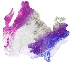
The new, advanced infrared (IR) imaging technique uses no chemical stains, instead scanning the sample with IR light to directly measure the chemical composition of the cells. The computer then translates spectral information from the microscope into chemical stain patterns, without the need to apply dyes to the cells.
"We're relying on the chemistry to generate the ground truth and act as the 'supervisor' for a supervised learning algorithm," says David Mayerich, first author of the study. Mayerich was a postdoctoral fellow at the Beckman Institute and now is a professor at the University of Houston. "One of the bottlenecks in automated pathology is the extensive processing that must be applied to stained images to correct for staining artifacts and inconsistencies. The ability to apply stains uniformly across multiple samples could make these initial image processing steps significantly easier and more robust."
The researchers reproduced a wide array of molecular stains by computationally isolating the spectra of specific molecules. This allows the user to simply tune to a required stain, for as many different stains as are necessary—all without damaging the original tissue sample, which can then be used for other tests.
"This approach promises to have immediate and long-term impact in changing pathology to a multiplexed molecular science—in both research and clinical practice," Bhargava says.
Full details of the work appear in the journal Technology; for more information, please visit http://dx.doi.org/10.1142/S2339547815200010.
-----
Follow us on Twitter, 'like' us on Facebook, connect with us on Google+, and join our group on LinkedIn