Optical refractive index of live cells could have use as molecular probes
Researchers at the Salk Institute for Biological Studies (La Jolla, CA) have discovered how to use the cephalopod proteins responsible for the instant color and iridescence changes displayed in octopuses, squid, and cuttlefish to manipulate the optical refractive index (RI) of mammalian cellular compartments. By changing this intrinsic physical property of the cell, microscopists could soon be using the RIs of live cells as molecular probes and capturing the resulting label-free quantitative phase images using Tomocube’s (Daejeon, South Korea) holotomography microscope.
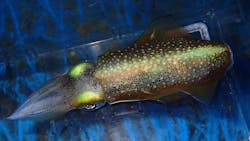
Gerald M. Pao and his colleagues set out to genetically manipulate the RI of living cells by synthesizing known cephalopod reflectin genes and cloning new reflectin genes expected to yield a higher RI from the wild Glitter or Bigfin reef squid (Sepioteuthis lessoniana). Epitope tagged for antibody recognition with either an HA or FLAG tag, with an added preprotrypsin signal sequence to target the protein into the secretory pathway, a C-terminal KDEL ER retention signal was added to specifically target the endoplasmic reticulum (ER) of cells. Their success is demonstrated in the quantitative phase contrast microscopy studies, where the Tomocube holotomography microscope measured an RI of up to 1.4—well above the ER RI of 1.35.
“Microscopists will tell you that RI is an intrinsic physical property of any material, including cellular components, and is not generally considered amenable to manipulation,” explains Aubrey Lambert, Tomocube’s Chief Marketing Officer. This work, he says, ends this notion and opens up a new era of control of the RIs of live cells, as the researchers have shown how to turn the cell itself into a molecular probe for quantitative phase imaging. “One use of this new technology may be to manipulate the RI to achieve transparency of scattering tissues through refractive index matching. Until now, this has required fixed, permeabilized whole organ samples, which precludes live imaging. However, with a genetically encoded protein, this may be conceptually possible,” Lambert says.
Reflectins proteins are the main components responsible for the structural coloration and iridescence in cephalopods, mainly used for camouflage. They are found in specialized cells, the iridophores, forming Bragg mirrors by alternating layers of high- and low-refractive-index material. Within the membrane stacks, reflectins form nanoparticles of <5 to >500 nm and confer the high optical refractive index property to iridophores. Reflectins are also utilized by cephalopods for white light scattering and to achieve chromatic invariance of chromatophores, pigment sacs, as these expand or contract.The Tomocube holotomography microscope delivers quantitative, nanoscale, real-time, and label-free 3D images of individual living cells quickly without any sample preparation. The holotomography images also deliver vital information on unique cell properties, including cell volume, shapes of subcellular organelles, cytoplasmic density, surface area, and deformability. The latest HT-2 model combines the quantitative phase imaging approach of label-free, 3D RI tomography with 3D fluorescence imaging.
Full details of the work are available at bioRxiv.
Source: Tomocube press release