Fluorescence monitors how protein aggregate present in Alzheimer's grows
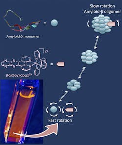
Researchers at Rice University (Houston, TX) have found a way to track the formation of soluble amyloid beta peptide aggregates implicated in the onset of Alzheimer's disease. The researchers, led by Angel Martí, Associate Professor of Chemistry, Bioengineering, and MSNE at Rice University, reported their development of a ruthenium-based fluorescent complex that binds to soluble, oligomeric amyloid beta peptides. As the peptides come together to form the large biomolecules called oligomers, the fluorescent additive binds and labels them. This enables the researchers to track the progress and movements of aggregates as they grow over time.
Amyloid plaques have long been suspected as the root cause of Alzheimer's, but recent studies suggest that oligomers—floating molecules with repeating peptide units—do far more damage. "There's a view in the field that soluble oligomers are the main cause of neuronal degeneration, because these oligomers are toxic to neurons," says Martí. "These oligomers are definitely associated with Alzheimer's pathology, so there's been a need for tools to help us study them."Oligomers are "virtually invisible" to Thioflavin T dyes commonly used to tag amyloid fibrils in lab studies, explains Martí. The ruthenium complexes solve that problem, as they take advantage of fluorescence anisotropy, in which the fluorescent response is polarized, glowing brighter in some directions than others. "It's a very old technique related to the rotation of molecules," he says. "When the molecule is in solution, it moves and is constantly rotating. When it's very small, it rotates very fast and the anisotropy is nearly zero. But when the same probe binds to a big macromolecule, it rotates more slowly. That's how we know we have oligomers, and then we can track their growth and propagation."
Lab tests showed oligomers forming in solution at different temperatures over hours. Martí notes that cold solutions slow the process, but at body temperatures, oligomers form "very fast and in large amounts. The speed at which they form at physiological temperatures is remarkable."
The lab also used its probes to see how neuroblastoma cells were affected in real time when injected with amyloid beta peptides. They revealed only 60% of cells injected with oligomers remained viable, while those treated with amyloid fibrils and monomers had higher viability, about 80%, suggesting the oligomers are indeed toxic, Martí says.For now, the ruthenium probes are meant for use only in the laboratory, Martí says. "It will be difficult to use these in the brain because there's too much scattering of light," he says. "They are made to take advantage of polarized light, and scattering would dampen that. But as a lab tool, they will allow researchers around the world to test the effects of other molecules on the rate of oligomer formation, and that's a big deal, They can quickly see if a drug delays or halts the formation of oligomers."
Full details of the work appear in the Journal of the American Chemical Society.


