Scanning fiber endoscope monitors carotid artery to track cardiovascular event risk
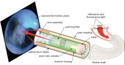
Knowing that strokes and heart attacks often strike without warning, a team of researchers at Michigan Medicine (University of Michigan; Ann Arbor, MI) has applied a previously developed scanning fiber endoscope (SFE) device to improve views of the carotid artery. The work could someday help physicians know who is at risk for a cardiovascular event by providing a better view of potential problem areas.
Related: Camera in a pill obtains high-resolution images inside the body
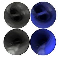
First author Luis Savastano, MD, a Michigan Medicine resident neurosurgeon, explains that the SFE allows them to see—in very high resolution—the surface of the vessels and any lesions, such as a ruptured plaque, that could cause a stroke. The technology, he says, may even be able to show which silent, but at-risk, plaques may cause a cardiovascular event in the future.
The SFE used in the study was invented and developed by co-author and University of Washington mechanical engineering research professor Eric Seibel, Ph.D. He originally designed it for early cancer detection by clearly imaging cancer cells that are currently invisible with clinical endoscopes.
The Michigan Medicine team used the SFE device to acquire high-quality images of possible stroke-causing regions of the carotid artery that may not be detected with conventional radiological techniques. They worked with senior author Thomas Wang, MD, Ph.D., professor of internal medicine, biomedical engineering, and mechanical engineering, and the H Marvin Pollard Collegiate Professor of Endoscopy Research.
The researchers generated images of human arteries using the SFE, which illuminates tissues with multiple laser beams, and digitally reconstructs high-definition images to determine the severity of atherosclerosis and other qualities of the vessel wall. It also uses fluorescence indicators to show key biological features associated with increased risk of stroke and heart attacks in the future.
An additional utility for the SFE is that it can also assist neurosurgeons with therapeutic interventions by guiding stent placement, releasing drugs and biomaterials and helping with surgeries, Seibel says.
Full details of the work appear in the journal Nature Biomedical Engineering; for more information, please visit http://dx.doi.org/10.1038/s41551-016-0023.


