Noninvasive multiphoton microscopy method predicts cancer malignancy
Scientists at Osaka University in Japan have shown that non-labeling multiphoton microscopy (NL-MPM) can be used for quantitative imaging of cancer that is safe and requires no resection, fixation, or staining of tissues. The work is expected to simplify and reduce the time it takes to diagnose cancer.
Related: Multiphoton microscopy detects skin cancers noninvasively
To diagnose cancer and determine the proper treatment plan, pathologists depend on biopsies from the patient. The cutting, fixing, and staining of the specimen generally provides reliable information, but these pathology-based diagnostic procedures require substantial time and cause tissue damage. Professor Masaru Ishii and his team of scientists have been exploring non-labeling microscopy methods as a means to observe and diagnose the cancer while preserving as much information as possible.
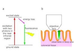
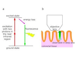
"MPM is an effective tool that enables the visualization of deep regions within living tissues and organs. We are developing NL-MPM to diagnose the malignancy of colorectal cancer," Ishii explains.
Colorectal cancer has some ideal properties for the application of NL-MPM. Traditionally, microscopy of living organisms has depended on the attachment of a fluorescent dye to the target tissue. However, these dyes are often toxic, and it would be desirable to visualize human tissues without the need for additional labeling. Colorectal cancer particularly affects epithelial tissue, which emanates a sufficient signal in NL-MPM without the addition of any foreign dye, because chemicals natural to the tissue will emit autofluorescence.
"MPM relies on second-harmonic generation (SHG) in addition to autofluorescence, and the basement membrane beneath the epithelial cells gives a strong SHG signal," Ishii explains, adding that the signal comes specifically from the basement membrane of the tissue, a region that is difficult to observe using contemporary staining techniques.
Using NL-MPM, Ishii's group, in collaboration with other scientists at Osaka University, was able to quantify the severity of the cancer using two parameters they created, index N and index S. Index N is a number that describes the size of the nuclei in the cell, while index S is a number that summarizes the intensity of SHG signals. Normal tissue was defined as that with an index N £ 9.5 (a larger index N indicating larger nuclei) and an index S > 3.1 (healthy tissue, because of its regular structure, gives off a stronger SHG signal).
"Any tissues that were outside these boundaries were deemed cancerous," Ishii says. The two indices also provide a highly reliable prediction of the malignancy of the cancer.
Ishii acknowledges that the technology needs improvement before it reaches clinical use, but is excited about its prospect for patient care. "We envision the use of endoscopy with the MPM system, as well as the use of NL-MPM during operations because of its rapid analysis," he says.
Full details of the work appear in the journal Scientific Reports.


