Noninvasive photoacoustic imaging method detects gum disease
Knowing that the conventional method to assess gum health can be invasive for the patient, engineers at the University of California San Diego have developed a dental imaging method that harnesses photoacoustic ultrasound to examine a patient's gums noninvasively.
Related: Photoacoustic microscopy enters the mainstream
Dentists currently use an instrument called a periodontal probe—a thin, hook-like metal tool that is marked like a tiny measuring stick and inserted in between the teeth and gums to see whether and how much the gums have shrunk back from the teeth, creating pockets. A pocket depth measuring 1–2 mm indicates healthy gums, while 3 mm and deeper is a sign of gum disease. Not only are procedures using a periodontal probe invasive, uncomfortable, and sometimes painful for the patient, measurements can also vary greatly between dentists, and the probe is only capable of measuring the pocket depth of one spot at a time.
So, Jesse Jokerst, a nanoengineering professor at UC San Diego and senior author of the study, worked with his research team to introduce a photoacoustic imaging method that can image the entire pocket depth around the teeth without requiring any painful poking and prodding.
The method begins by rinsing the mouth with a paste made of commercially available food-grade squid ink mixed with water and cornstarch. The squid-ink-based rinse serves as a contrast agent for the photoacoustic imaging method, which involves shining a light signal—usually a short laser pulse—onto a sample,which heats up and expands to generate an acoustic signal that researchers can analyze.
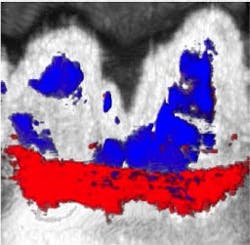
Squid ink naturally contains melanin nanoparticles, which absorb light. During the oral rinse, the melanin nanoparticles get trapped in the pockets between the teeth and gums. When researchers shine a laser light onto the area, the squid ink heats up and quickly swells, creating pressure differences in the gum pockets that can be detected using ultrasound. This method enables researchers to create a full map of the pocket depth around each tooth—a significant improvement over the conventional method.
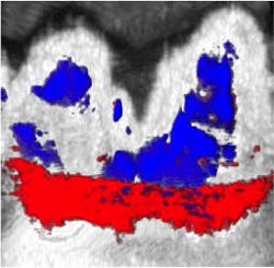
Researchers tested their photoacoustic imaging method in a pig model containing a mix of shallow and deep pockets in the gums. While their results closely matched measurements taken using a periodontal probe, they were also consistent across multiple tests. On the other hand, measurements with the periodontal probe varied significantly from one test to another.
Moving forward, the team will be collaborating with dentists and testing their method in humans. Future work also includes minimizing the bitter, salty taste of the squid ink oral rinse, and replacing laser lights with inexpensive, more portable light systems like LEDs. The team's ultimate goal is to create a mouthpiece that uses this technology to measure periodontal health.
Full details of the work appear in the Journal of Dental Research.