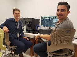
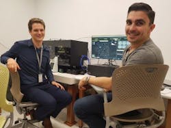
Translational Research Institute acquires Livecyte cell imaging and analysis system
The Translational Research Institute (TRI; Brisbane, Australia), which was formed to accelerate application of innovative scientific research to improve healthcare outcomes, has adopted Phasefocus' (Sheffield, England) Livecyte cell imaging and analysis system.
Related: Ptychography method uses phase information for high-contrast, label-free imaging
The Livecyte system enables quantitative, label-free, live-cell imaging and analysis of single and multiple cell types in heterogeneous cell populations using ptychographic quantitative phase imaging (QPI). Requiring only low-level illumination, the system provides a gentle experimental environment that mimics the behavior of cells in real life, allowing its use in more clinically and physiologically relevant primary and stem cell populations alongside traditional cell assays.
Professor Brian Gabrielli, who leads the Melanoma Research Group at TRI, saw the capabilities of Livecyte during a visit to the University of York (England) and immediately recognized its potential to gain a better understanding of cell cycle defects associated with melanoma. Following a review of the system's functionality, the research team received funding to procure a system for the Microscopy core facility at TRI.
For more information, please visit www.phasefocus.com.

