Potassium-sensitive optical nanosensor improves brain mapping accuracy
Researchers at the University of Lausanne (Switzerland) have developed an optical nanosensor that enables less invasive, more accurate measurement and spatiotemporal mapping of the brain. The development could someday result in design of future multimodal sensors and have utility in a broader range of applications.
Related: Advanced light microscopy enables rapid mapping of brain structure and function in high resolution
Neuronal activity results in the release of ionized potassium into extracellular space. Under active physiological and pathological conditions, elevated levels of potassium need to be quickly regulated to enable subsequent activity. This involves diffusion of potassium across extracellular space as well as re-uptake by neurons and astrocytes.
Measuring levels of potassium released during neural activity has involved potassium-sensitive microelectrodes, and to date has provided only single-point measurement and undefined spatial resolution in the extracellular space.
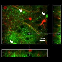
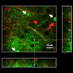
With a fluorescence-imaging-based, ionized-potassium-sensitive nanosensor design, the research team was able to overcome challenges such as sensitivity to small movements or drift and diffusion of dyes within the studied region, improving accuracy and enabling access to previously inaccessible areas of the brain.
This potassium-sensitive nanosensor is likely to aid future investigations of chemical mechanisms and their interactions within the brain, the researchers say. The spatiotemporal imaging created by collected data will also allow for investigation into the possible existence of potassium micro-domains around activated neurons and the spatial extent of these domains. The study confirms the practicality of the nanosensor for imaging in the extracellular space, and also highlights the range of possible extensions and applications of the nanosensor strategy.
Full details of the work appear in the journal Neurophotonics; for more information, please visit http://dx.doi.org/10.1117/1.nph.4.1.015002.

