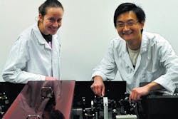
New microscope provides live-cell imaging breakthrough for cancer treatment and drug discovery
Glasgow, Scotland-- A new microscope has been designed and built at the Centre for Biophotonics of the Strathclyde Institute of Pharmacy and Biomedical Sciences. Designed and built by Dr. Gail McConnell, Reader and RCUK Academic Fellow, and post-doctoral researcher Dr. Wei Zhang, the microscope uses a less-complex laser system than previously available for obtaining crucial, highly detailed images of cells, tissues and drugs.
Extending a known microscopy technique, the new imaging platform adopts a simpler, less expensive approach that uses only one laser, and the levels of light involved are sufficiently low to avoid causing cell damage. The microscope development was funded by the Biotechnology and Biological Sciences Research Council as part of its Technology Development Research Initiative program in 2007.
The new microscope eliminates the need to add often-toxic fluorescent labeling normally required to allow scientists to see specific areas of interest within material, reducing the likelihood of the material becoming disrupted. Using the new microscope system, coupled with the chemical properties of the materials themselves, scientists can obtain high-definition, 3-D images.
"By producing chemically specific images of cells and tissue without adding potentially disruptive dyes, life sciences researchers can visualize sub-cellular and cellular structures in three dimensions and with minimum intervention. This opens up new vistas in live cell imaging and provides biomedical researchers with the unparalleled ability to study biological function, which provides a unique insight into the fundamental spark of life," commented Dr. McConnell.
