Researcher combines AFM and FRET to study single biological molecules
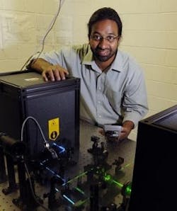
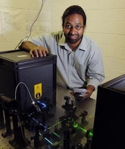
Sanjeevi Sivasankar, an Iowa State University assistant professor of physics and astronomy and an associate of the U.S. Department of Energy's Ames Laboratory, came up with the idea of developing and building a unique, single-molecule microscope. Because different kinds of cells have different kinds of surface proteins (cadherins), the typical tools for observing and measuring those proteins focus on tens of thousands of them at a time—providing data on the average molecule in a sample, but not on a single molecule.
The new idea was to combine two single-molecule technologies that had been used separately: atomic force microscopy (AFM), which manipulates molecules and measures forces; and fluorescence resonance energy transfer (FRET), which observes single molecules at very high resolution. Sivasankar, a post-doctoral researcher at Stanford University and the University of California-Berkeley, who worked with Steven Chu, the current U.S. Secretary of Energy and co-winner of the 1997 Nobel Prize in Physics, hopes that the combined instrument will advance studies in biomedical research, drug discovery, cancer diagnostics and biosensing applications.
Sivasankar brought the idea for an integrated, single-molecule instrument to Ames when he started at Iowa State and the Ames Laboratory in 2008. He's since built a laboratory prototype and improved its measurement capabilities and efficiency.
Sivasankar and his research group—Iowa State and Ames Laboratory post-doctoral researchers Hui Li and Sabyasachi Rakshit, plus Iowa State doctoral students Kristine Manibog and Chi-Fu Yen—spend about half their time developing the new instrument. The other half is spent on studies of single molecule biophysics.
The new instrument, for example, is advancing studies of cadherins and DNA, Sivasankar said. The researchers are also using it to study semiconducting nanocrystals in a research collaboration with Paul Alivisatos, the director of the Lawrence Berkeley National Laboratory in Berkeley, CA.
Sivasankar said he expects discoveries based on data from the new microscope will soon be published in scientific journals.
As he tests and proves the instrument, Sivasankar will begin working with Novascan Technologies Inc. of Ames to continue development. Earlier this year, he won a $120,075 grant from the Grow Iowa Values Fund, a state economic development program, to support commercialization of the microscope. University startup funds and an award from the March of Dimes have also supported development of the microscope.
Novascan Technologies is a nanotechnology company with operations in the Iowa State University Research Park and downtown Ames. It was founded by Raj Lartius, a former Iowa State student and faculty member who's now the company's chief executive officer. For 12 years, it has developed and globally marketed single molecule instrumentation based on AFM.
Sivasankar said the immediate goal is to transform the microscope from its bulky, prototype stage to an instrument that's novel, compact, easy to use and can be manufactured at a competitive price.
-----
Posted by Lee Mather
Subscribe now to BioOptics World magazine; it's free!

