Microscopy method images whole brains at nanoscale, and rapidly
A team of researchers at the Massachusetts Institute of Technology (MIT; Cambridge, MA), in collaboration with the Howard Hughes Medical Institute's Janelia Research Campus (Ashburn, VA), has developed a technique that combines expansion microscopy with lattice light-sheet microscopy. The pairing enables nanoscale imaging of fly and mouse neuronal circuits and their molecular constituents, and is shown to be approximately 1000X faster than other methods.
Scientists Ruixuan Gao and Shoh Asano, who are a part of Ed Boyden's lab at MIT, developed expansion microscopy, which works well on single cells or thin tissue sections imaged in conventional light microscopes. They wanted, however, to image vastly larger chunks of tissue, with the ability to see complete neural circuits spanning millimeters or more.
To do so, the scientists needed a microscope that was high-speed, high-resolution, and relatively gentle. So, they turned to Eric Betzig at the Howard Hughes Medical Institute's Janelia Research Campus, whose team had used their lattice light-sheet microscope to image the rapid subcellular dynamics of sensitive living cells in 3D. Combining the two microscopy techniques could potentially offer rapid, detailed images of wide swaths of brain tissue.
Now, in their new collaboration, the two research teams have imaged the entire fruit fly brain and sections of mouse brain the thickness of the cortex. Their combined method offers high resolution with the ability to visualize any desired protein rapidly. Imaging the fly brain in multiple colors took 62.5 hours, compared to the years it would take using an electron microscope.
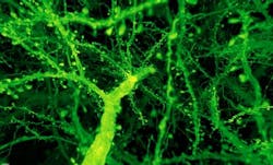
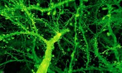
A forest of dendritic spines protrudes from the branches of neurons in the mouse cortex. (Image credit: Gao et al./Science 2019)
The lattice light-sheet microscope sweeps an ultrathin sheet of light through a specimen, illuminating only that part in the microscope's plane of focus. That helps out-of-focus areas stay dark, keeping a specimen's fluorescence from being extinguished.
Yoshinori Aso and the FlyLight team provided high-quality fly brain specimens, which Gao and Asano expanded and used to collect some-50,000 cubes of data across each brain. Those images required complicated computational stitching to put the pieces back together, work led by Stephan Saalfeld and Igor Pisarev.
After expanding the fruit fly brain to four times its usual size, scientists used lattice light-sheet microscopy to image all of the dopaminergic neurons (green). (Image credit: Gao et al./Science 2019)
Next, Srigokul Upadhyayula from Harvard Medical School (also in Cambridge, MA) and Boston Children's Hospital (Boston, MA), a co-first author of the report, analyzed the combined 200 terabytes of data and created movies that showcase the brain's intricacies in vivid color. He and his coauthors investigated more than 1500 dendritic spines, imaged fatty sheaths that insulate mouse nerve cells, highlighted all of the dopaminergic neurons, and counted all the synapses across the entire fly brain.
However, as with any kind of superresolution fluorescence microscopy, it can be hard to decorate proteins with enough fluorescent bulbs to see them clearly at high resolution, Betzig says. And since expansion microscopy requires many processing steps, there is still the potential for artifacts to be introduced. Because of this, he says, "we worked very hard to validate what we've done, and others would be well advised to do the same."
Now, Gao and the Janelia team are building a new lattice light-sheet microscope, which they plan to move to Boyden's lab at MIT. "Our hope is to rapidly make maps of entire nervous systems," Boyden says.
Full details of the work appear in the journal Science.


