Microscopy method peers into cell structures of living fish
Researchers from the Karlsruhe Institute of Technology (KIT; Karlsruhe, Germany), the Max Planck Institute for Polymer Research (Mainz, Germany), and the American National Institutes of Health (NIH; Bethesda, MD) have developed a new microscopy method to visualize cell structures measuring an eighth of a micrometer in size in living zebrafish larvae.
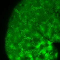
Zebrafish larvae, which are completely transparent, suit genetic cell study when injected with fluorescent dye, explains Marina Mione of KIT, who led the work. In this case, the researchers sought to visualize parts of the microtubuli (the cellular skeleton of fish), which measure around 100 µm in length and 20 nm in diameter.

Dubbed smart illumination, the researchers' microscopy method illuminates the object only at a certain spot rather than completely, minimizing scattered light and sharply representing illuminated detail. Taking a series of images at variable illumination and within a few seconds, the researchers then processed them via computer to obtain an overall image. Smart illumination also allowed the researchers to adjust the depth of field, image at various depth levels, and then produce a 3D image on the computer. Their method makes it possible to reach resolutions of 145 nm in the plane and 400 nm in between, says Mione.
The work has been published in Nature Methods; for more information, please visit http://www.nature.com/nmeth/journal/vaop/ncurrent/full/nmeth.2025.html.
-----
Follow us on Twitter, 'like' us on Facebook, and join our group on LinkedIn
Follow OptoIQ on your iPhone; download the free app here.
Subscribe now to BioOptics World magazine; it's free!