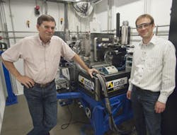
X-ray 'bionanoprobe' boosts study of cellular processes
A team of researchers from Northwestern University's Department of Radiation Oncology (Chicago, IL) and the U.S. Department of Energy's (DOE's) Argonne National Laboratory (Argonne, IL) have developed a non-destructive x-ray microscopy solution to image cryogenically preserved cells and advance studies of intracellular biology. The solution could be used to study and potentially treat cancer, neurological disorders, and other diseases and conditions involving the accumulation of metals within cells.
Dubbed the Bionanoprobe, which features a noninvasive 3D x-ray microscope (Xradia's UltraSPX; Pleasanton, CA), the team's solution delivers high-resolution x-ray trace element mapping and tomography of cryogenically preserved samples down to 30 nm—enabling researchers to uniquely understand what occurs inside cells, says Dr. Gayle Woloschak, Professor of Radiation Oncology at the Robert H. Lurie Comprehensive Cancer Center, Northwestern University Feinberg School of Medicine, and principal investivator on the project. Bionanoprobe could enable researchers to pinpoint what role trace metals like zinc or iron play in natural cellular processes like cell division and aging, for example, she says.
Deployed last fall by the Life Sciences Collaborative Access Team (LS-CAT) at Northwestern, Bionanoprobe is expected to produce new insights into the behavior of nanoparticles within cells for fields such as pharmacology and toxicology, says Dr. Keith Brister, LS-CAT operations manager. Bionanoprobe enables imaging in high-resolution x-ray fluorescence (XRF), transmission, spectroscopy, and tomography. Combining the techniques provides information on elemental content, structure, and chemical state, in 3D, over a wide range of length scales to examine cells and other samples at high resolutions.
"Using one technique makes it possible to compare elements more precisely," says Woloschak. "Traditionally, looking at tissue under a regular microscope then moving to an electron microscope requires that we use different sections and preparation techniques, which can introduce artifacts and make it hard to compare and co-localize features. The best we could do is match as closely as possible; we couldn't look at the exact item under varying conditions."
Bionanoprobe combines ultra-high-resolution trace element mapping with cryogenic sample preservation and tomographic capabilities, enabling study of cells and tissue in a state closely resembling that of being alive, while minimizing the effects of radiation damage that can distort the results. Tomography is needed to exactly localize the features of interest inside the cell.
Bionanoprobe's cryogenic sample-handling system allows researchers to move the same cryogenically preserved sample from the x-ray nanoprobe to a transmission x-ray microscope, or potentially other cryo instruments, says Dr. Wenbing Yun, founder and CTO of Xradia. "Scientists look at tissue down to subcellular locations with one technique, which is virtually impossible otherwise," he adds.
-----
Follow us on Twitter, 'like' us on Facebook, and join our group on LinkedIn
Follow OptoIQ on your iPhone; download the free app here.
Subscribe now to BioOptics World magazine; it's free!
