Handheld fluorescent probe assists in brain cancer surgery
The first of its kind to use fluorescence to distinguish cancerous tissue from normal tissue in low-grade brain tumors, researchers at the Norris Cotton Cancer Center and Dartmouth College's Thayer School of Engineering (both in Lebanon, NH) have developed a fluorescent probe that could help brain surgeons. The probe tool, now already in use at the Cancer Center for brain surgery, could someday be used for surgeries for a variety of cancers.
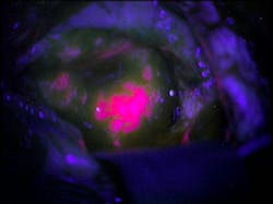
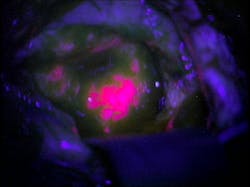
To improve their ability to differentiate between tumor cells and healthy tissue, surgeons can have patients take an oral dose of the chemical 5-aminolevulinic acid (ALA). An enzyme metabolizes ALA, producing the fluorescent protein protoporphyrin IX (PpIX). Tumor cells have a higher metabolic rate than normal cells, so they accumulate more PpIXâand therefore fluoresce when exposed to blue light.
But this method was thought to be not sensitive enough to highlight brain tumors that are less metabolically active, like low-grade gliomas. To address this problem, MD-Ph.D. student Pablo Valdes, Ph.D., and his research mentorsâDavid Roberts, MD, the chief of neurosurgery at Dartmouth-Hitchcock Medical Center, and Keith Paulsen, Ph.D., a professor of biomedical engineering at Thayer School of Engineering and a member of the Cancer Imaging and Radiobiology Research Program at Norris Cotton Cancer Centerâdeveloped a probe that combines violet-blue and white light to simultaneously analyze both the concentration of PpIX and four other tumor biomarkers: PpIX breakdown products, oxygen saturation, hemoglobin concentration, and irregularity of cell shape and size. The probe reads how light travels when it hits the tissue, sends this data to a computer, runs it through an algorithm, and produces a straightforward answer as to whether the tissue is cancerous.
The Thayer/Norris Cotton Cancer Center/Dartmouth-Hitchcock Medical Center team, which has been working on the brain probe for about six years in collaboration with researchers from the University of Toronto (Toronto, ON, Canada), built on research that was originally conducted in Germany about 15 years ago. But the German research did not focus on low-grade tumors. The assumption, says Paulsen, was that, due to the blood-brain barrier, low-grade tumors did not have enough PplX and therefore would not fluoresce.
In a pilot study, Roberts operated on 10 patients with gliomas. He used a microscope throughout the surgery to see the fluorescence and used the handheld probe to evaluate sections of the tissue where the fluorescence was not definitive. After the surgeries were complete, a pathologist evaluated how accurately the probe had identified tumor tissue.
Diagnoses based on fluorescence only had an accuracy of 64 percent. But when Roberts used the probe, the accuracy increased to 94 percent, meaning that Roberts was much more successful in differentiating between tumor tissue and normal tissue. Although the study was small, it introduces a promising method to help surgeons remove only what they want and nothing more.
Paulsen says that a protocol is in place for a similar study on lung cancer tumors. He thinks the brain probe may have applications for other cancers as well, but for now the research will have to proceed one cancer type at a time.
For more information on the work, which has been published in the Journal of Biomedical Optics, please visit http://spiedigitallibrary.org/jbo/resource/1/jbopfo/v16/i11/p116007_s1.
-----
Follow us on Twitter, 'like' us on Facebook, and join our group on LinkedIn
Laser Focus World has gone mobile: Get all of the mobile-friendly options here.
Subscribe now to BioOptics World magazine; it's free!