Fluorescence imaging technique visualizes blood flow in living animals

Scientists at Stanford University (Stanford, CA) have developed a fluorescence imaging technique for watching blood flow in living animals. It involves carbon nanotubes and lasers, and will allow researchers to better study arterial diseases and therapies.
The fluorescence technique, called near infrared-II imaging (NIR-II), involves first injecting water-soluble carbon nanotubes into the living subject's bloodstream. The researchers then shine a laser with NIR light (a wavelength of about 0.8 µm) over the subject; in this case, a mouse. The light causes the specially designed nanotubes to fluoresce at a longer wavelength of 1â1.4 µm, which is then detected to determine the blood vessels' structure.
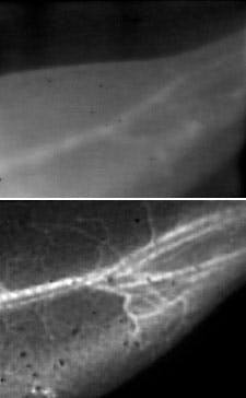
The fact that the nanotubes fluoresce at substantially longer wavelengths than conventional imaging techniques is critical in achieving the stunningly clear images of the tiny blood vessels: longer wavelength light scatters less, and thus creates sharper images of the vessels. Another benefit of detecting such long wavelength light is that the detector registers less background noise since the body does not does not produce autofluorescence in this wavelength range.
In addition to providing fine details, the techniqueâdeveloped by Stanford scientists Hongjie Dai, professor of chemistry; John Cooke, professor of cardiovascular medicine; and Ngan Huang, acting assistant professor of cardiothoracic surgeryâhas a fast image acquisition rate, allowing researchers to measure blood flow in near real time.
The ability to obtain both blood flow information and blood vessel clarity was not previously possible, and will be particularly useful in studying animal models of arterial disease, such as how blood flow is affected by the arterial blockages and constrictions that cause, among other things, strokes and heart attacks.
"For medical research, it's a very nice tool for looking at features in small animals," Dai says. "It will help us better understand some vasculature diseases and how they respond to therapy, and how we might devise better treatments."
Because NIR-II can only penetrate a centimeter, at most, into the body, it won't replace other imaging techniques for humans, but it will be a powerful method for studying animal models by replacing or complementing x-ray, CT, MRI, and laser Doppler techniques.
The next step for the research, and one that will make the technology more easily accepted for use in humans, is to explore alternative fluorescent molecules, Dai says. "We'd like to find something smaller than the carbon nanotubes but that emit light at the same long wavelength, so that they can be easily excreted from the body and we can eliminate any toxicity concerns."
The work was published online in Nature Medicine; for more information, please visit http://www.nature.com/nm/journal/vaop/ncurrent/full/nm.2995.html.
-----
Follow us on Twitter, 'like' us on Facebook, and join our group on LinkedIn
Laser Focus World has gone mobile: Get all of the mobile-friendly options here.
Subscribe now to BioOptics World magazine; it's free!