Research microscope donation helps spur minority students' interest in science
Recognizing that minority students do not tend to choose science as a career path, Emil Bogenmann, Ph.D., Ed.D., director for Research Education at the Saban Research Institute of Children’s Hospital (Los Angeles, CA), established the Latino & African American High School Internship Program (LA-HIP), a biomedical summer research and college preparatory program for senior high school students who attend school in South or East Los Angeles. Established in 2005, interns in the program work for six weeks in medical research labs performing hands-on experiments relevant to childhood diseases. What's more, the program recently received a donation of research microscopes from Carl Zeiss (Oberkochen, Germany) to help motivate the students further.
The LA-HIP program begins with a laboratory training course, during which the 16 interns work in small groups to learn the fundamentals of laboratory experimentation, note taking and conduct. After this introduction, they perform five weeks of hands-on research in the laboratories of principal investigators at the Saban Research Institute. Their research focuses on the biology of infectious agents and their mechanisms of actions, metastasis and invasive behavior of cancer cells, lung development, organ injury and repair, and zebrafish heart regeneration, among other topics.
Students work full-time, performing sophisticated experiments normally not learned until graduate school. The internship concludes with a science symposium at which all LA-HIP interns present their research accomplishments to guests, families, hospital leadership, trustees, scientists, and mentors.
Roxana Rodriguez, a senior at Bravo Medical Magnet High School, worked in the laboratory of Dr. Bouret studying neuronal circuitry formation during early mouse brain development. Staining brain slices with fluorescent antibodies and viewing them with a Zeiss confocal microscope exposed her to the cutting edge of microscopy. Roxana was so enthusiastic about her work that she declared that her career will involve research and medicine, saying, “LA-HIP helped me decide that I still want a career in science and in medicine in the future. Overall, LA-HIP has been a pivotal point in my life that will help me reach my current and long-term goals.”
Similarly, Jackie Hernandez, a senior at Aspire High School, and her lab partner, Stephanie Leyva, a senior at the California Academy of Math and Science (CAMS), obtained intriguing results in their studies of brain invasion by bacteria. They demonstrated that avian specific bacterial pathogens are equally if not more invasive into human brain endothelial cells than their human counterpart.
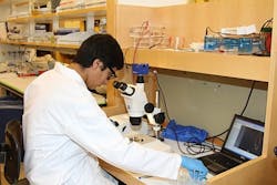
Carl Zeiss supplied a Stemi 2000C stereo microscope with an AxioCam ERc 5s camera, which can also work as a stand-alone imaging station. Mark Mobilia, a local sales representative for Carl Zeiss Microscopy, has worked to install the equipment and provide technical support during the course of the program.
Bogenmann created an experiment using planaria worms (flat worms) to illustrate regeneration: Students would begin by observing the worm under the microscope, then amputate the head from the body, photograph it again, leave it for a few days, and then observe the worm under the microscope a few days later to find that both the trunk and the head regenerated new worms.
“Students come here in the first week to learn laboratory fundamentals so they can learn how to isolate DNA and look at microscopic cells and tissue, but many biological concepts such as stem cells and their potentials are difficult to envision,” says Dr. Bogenmann. “So this year I conceived this experiment as a way for students to personally experience the regenerative process of stem cells, as they observed pieces of the planaria regenerating into new planaria with head and tail.”
The availability of the microscope and digital camera hooked to a laptop enabled the students to take pictures and make observations on their own specimens. They also collaborated with Alejandro Sánchez Alvarado, Ph.D., and his laboratory at the Stowers Institute for Medical Research (Kansas City, MO)—planaria experts who provided them with siRNA molecules for one gene extremely important to head regeneration. Feeding these siRNA molecules before amputation created new planaria that had structure on both ends of the worm, thus demonstrating the power of gene expression during the regrenerative process.
Thanks to the success of the planaria experiment, Bogenmann is already strategizing about spending more time on the experiment next year before the students head off to their research labs. He hopes to obtain even more sophisticated camera equipment so the interns can record short videos of the different stages of the planaria regenerative process.
“The students showed great intelligence and maturity, the experiment worked perfectly, and it was an excellent way to get kids motivated about science,” notes Mobilia. “Carl Zeiss is fully committed to helping recruit more minorities into science. We have already made plans to supply equipment for next year’s crop of interns, and even hope to increase our financial and technical support.”
-----
Follow us on Twitter, 'like' us on Facebook, and join our group on LinkedIn
Laser Focus World has gone mobile: Get all of the mobile-friendly options here.
Subscribe now to BioOptics World magazine; it's free!