Spectroscopy boosts photoluminescent probes to study DNA sequences
Rice University (Houston, TX) researchers have optimized the sensitivity of photoluminescent probes using time-resolved spectroscopy, yielding results nearly twice as good as fluorescence spectroscopy does when probing for specific DNA sequences.
Led by Angel Martí, an assistant professor of chemistry and bioengineering, along with co-author Kewei Huang, a graduate student in Martí's group, their spectroscopic technique involves a luminescent probe called a molecular beacon, which is designed to attach to a target like a DNA sequence and then light up.
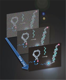
Time-resolved spectroscopy is like taking a film instead of a snapshot, says Martí. "We create a kind of movie that allows us to see a specific moment in the process where photoluminescence is occurring. Then, we can filter out the shadows that obscure the measurement or spectra we're looking for," he says.
To prove their method, Martí and Huang tested ruthenium- and iridium-based light-switching probes under standard fluorescent and time-resolved spectroscopy. The hairpin-shaped probes' middles are designed to attach to a specific DNA sequence, while the ends are of opposite natures. One carries the fluorophore (iridium or ruthenium), the other a chemical quencher that keeps the fluorescence in check until the probe latches onto the DNA. When that happens, the fluorophore and the quencher are pulled apart and the probe lights up.
In combination with a technique called fluorescence lifetime microscopy (FLIM), the Rice calculations may improve results from other diagnostic tools that gather data over time, such as magnetic resonance imaging (MRI) machines used by hospitals.
The work appears in the journal Analytical Chemistry; for more information, please visit http://pubs.acs.org/doi/abs/10.1021/ac3019894.
