Engineered DNA barcode boosts fluorescence microscopy potential

In efforts to develop a better molecular tool for fluorescence microscopy, researchers at the Wyss Institute for Biologically Inspired Engineering at Harvard University (Cambridge, MA) have created a new kind of barcode that could be available in almost any style to enable scientists to gather much more vital information than previously able. The DNA barcode they've created harnesses the natural ability of DNA to self-assemble.
Currently, scientists use fluorescence microscopy to pair fluorescent elements with molecules they know will attach to the part of the cells they want to investigate. Illuminating the sample triggers each kind of element to fluoresce at a particular wavelength of lightâsuch as red, blue, or greenâindicating where the molecules of interest are.
However, fluorescence microscopy is limited by the number of colors availableâthree or fourâand sometimes the colors get blurry. But the Wyss researchers' DNA barcode tool enables colored dots to be arranged into geometric patterns or fluorescent linear barcodes, and the combinations are almost limitlessâsubstantially increasing the number of distinct molecules or cells scientists can observe in a sample, and the colors are easy to distinguish.
In the researchers' method, DNA origami follows the basic principles of the double helix in which the molecular bases A (adenosine) only bind to T (thymine), and C (cytosine) bases only bind to G (guanine). With those "givens" in place, a long strand of DNA is programmed to self-assemble by folding in on itself with the help of shorter strands to create predetermined forms--much like a single sheet of paper is folded to create a variety of designs.
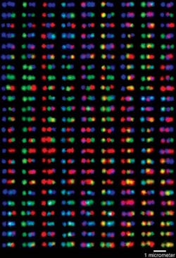
To these more structurally complex DNA nanostructures, researchers can then attach fluorescent molecules to the desired spots, and use origami technology to generate a large pool of barcodes out of only a few fluorescent molecules. That could add a lot to the cellular imaging "toolbox" because it enables scientists to potentially light up more cellular structures than ever possible before.
"The intrinsic rigidity of the engineered DNA nanostructures is this method's greatest advantage; it holds the fluorescent pattern in place without the use of external forces. It also holds great promise for using the method to study cells in their native environments," explains Peng Yin, Wyss core faculty member and study co-author. As proof of concept, the research team demonstrated that one of their new barcodes successfully attached to the surface of a yeast cell.
The researchers still need to determine what happens when each of the fluorescent barcodes are mixed together in a cell sample, which is routine in real-life biological and medical imaging systemsâbut it's low-cost, easy to do, and more robust compared to current methods, says Yin.
Potential applications for their DNA barcode method include targeted drug-delivery mechanisms and the ability to improve upon observation of cellular and molecular activities at a disease site using the latest medical imaging techniques.
The work appears in Nature Chemistry; for more information, please visit http://www.nature.com/doifinder/10.1038/nchem.1451.
-----
Follow us on Twitter, 'like' us on Facebook, and join our group on LinkedIn
Laser Focus World has gone mobile: Get all of the mobile-friendly options here.
Subscribe now to BioOptics World magazine; it's free!