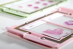
Extra-large slide scanning by Leica Microsystems
The SCN400 and SCN400 line scanners from Leica Microsystems (Wetzlar, Germany) offer an extra-large capture area for digital pathology of large tissue sections. For applications including neuroscience and toxicological pathology, the scanners capture extra-large jumbo (113 × 76 mm), double (52 × 76 mm), and traditional (26 × 76 mm) slides in either brightfield or fluorescence.
-----
PRESS RELEASE
Leica Microsystems Introduces Extra Large Slide Scanning for Digital Pathology at Neuroscience
Double and Jumbo Slide Scanning on the Leica SCN400
Wetzlar, Germany. The largest capture area of any line scanner in digital pathology is now possible with the Leica SCN400 and SCN400 F. The latest release in Leica’s Total Digital Pathology portfolio enables the digitization of very large specimens, not possible with any other line scanning digital pathology system.
This development provides a new level of flexibility in digital pathology, enabling users to scan a wide range of samples on a single system. Traditional (26 x 76 mm), double (52 x 76 mm) and now extra-large Jumbo (113 x 76 mm) slides can all be captured in either brightfield or fluorescence using the Leica SCN400 and SCN400 F scanners.
Existing users of the Leica SCN400 digital slide scanner range can benefit from being able to implement large slide scanning, without any hardware changes or physical updates to their scanners.
To view some sample large slide scans, please visit our Virtual Slide Gallery at www.leica-microsystems.com/products/digital-pathology.
Dr. Donal O’Shea, Head of Digital Pathology in Leica Microsystems says, “Line scanning in digital pathology has, until now, been limited to single and double slides. The introduction of extra-large slide scanning is a significant advancement for neuroscience, toxicological pathology and anyone dealing with large tissue sections. The cost and time saving benefits of sharing large tissue sections digitally rather than physically is a valuable tool for our users.”
Leica Microsystems’ Total Digital Pathology portfolio scanning range enables the capture of the widest range of sample types whether brightfield, fluorescence or FISH demonstrating Leica’s drive and innovation in this market. Leica Microsystems’ broad range of interoperable Digital Pathology components provides customers with the flexibility to choose the solution that is right for their needs.
Experience Leica Microsystems’ Total Digital Pathology solutions at booth 2825 during the Society for Neuroscience annual meeting, from November 12-16 in Washington, DC.
Leica Microsystems is a world leader in microscopes and scientific instruments. Founded as a family business in the nineteenth century, the company’s history was marked by unparalleled innovation on its way to becoming a global enterprise.
Its historically close cooperation with the scientific community is the key to Leica Microsystems’ tradition of innovation, which draws on users’ ideas and creates solutions tailored to their requirements. At the global level, Leica Microsystems is organized in four divisions, all of which are among the leaders in their respective fields: the Life Science Division, Industry Division, Biosystems Division and Medical Division.
Leica Microsystems’ Biosystems Division, also known as Leica Biosystems, offers histopathology laboratories the most extensive product range with appropriate products for each work step in histology and for a high level of productivity in the working processes of the entire laboratory.
The company is represented in over 100 countries with 12 manufacturing facilities in 7 countries, sales and service organizations in 19 countries and an international network of dealers. The company is headquartered in Wetzlar, Germany.
