Fluorescence approach detects drug-resistant tuberculosis in living bacilli
Scientists at the Antwerp Institute of Tropical Medicine (ITG; Antwerp, Belgium), using an updated approach to a fluorescence technique, have succeeded in detecting drug-resistant tuberculosis (TB) in living bacilli. What's more, their technique enables detection in resource-limited locations.
Viewing sputum smears under a microscope is still the gold standard for TB screening, but it cannot differentiate between living and dead bacilli. And even though polymerase chain reaction (PCR) technology can determine whether or not the bacillus is from a resistant strain, it is unfeasible in resource-limited areas due to its cost. It also is impossible to cultivate every sample and then target it with every possible antibiotic to survey which ones still work for an individual patient.
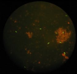
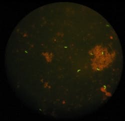
Recognizing these limitations, lead scientist Armand Van Deun and colleagues used vital staining with fluorescein diacetate (FDA) to only stain living TB bacilli in a sputum smear, enabling them to immediately see those bacilli escaping treatment. And they improved the detection of the luminous bacilli by replacing a fluorescence microscope with an LED illumination microscope.
The scientists' test enables detection of a high number of resistant TB bacilli that otherwise would have been discovered too late or not at all. They report that three times more patients could directly switch to the correct second-line treatment without losing time on a regimen ineffective against their resistant bacilli. And their technique can cut in half the number of cases where doctors start a retreatment because it ascertains that the bacilli detected by fluorescence microscopy in fact are dead ones, which do not require further treatment.
Together with colleagues in Bangladesh, the scientists tested their approach in the field for four years, thanks to a grant made possible by the Damien Foundation.
For more information on their workâwhich appears in The International Journal of Tuberculosis and Lung Diseaseâplease visit http://dx.doi.org/10.5588/ijtld.11.0166.
-----
Follow us on Twitter, 'like' us on Facebook, and join our group on LinkedIn
Laser Focus World has gone mobile: Get all of the mobile-friendly options here.
Subscribe now to BioOptics World magazine; it's free!

