Fluorescent imaging reveals cell membrane repair mechanism
Using high-resolution fluorescent imaging, scientists at the Karlsruhe Institute of Technology (KIT; Karlsruhe, Germany) and Heidelberg University (Heidelberg, Germany) have observed cell membrane repair in real time in a living organism for the first time.
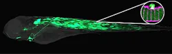
The researchers tagged repair proteins with fluorescent proteins in muscle of the transparent zebrafish larvae; then, with a laser, they burned tiny holes into the plasma membrane of muscle cells and followed the repair of the holes under the microscope. Doing so enabled them to show that membrane vesicles—together with two proteins Dysferlin and Annexin A6—rapidly form a repair patch; other Annexins accumulate subsequently on the injured membrane.
The studies suggest that the cell assembles a multilayered repair patch from the inside that seals off the cell's interior from the extracellular environment. What's more, they found that there is a specialized membrane area that quickly supplies the membrane that is needed for sealing the plasma membrane hole.
This animal model for membrane repair will contribute to the identification of new proteins in this sealing process and will help elucidate the underlying mechanisms. The results may contribute to the development of therapies for human myopathies and open up new possibilities in biotechnology.
The results of the work have published in Developmental Cell: http://www.sciencedirect.com/science/article/pii/S1534580711005740.