Optical microscopy technique enables detailed insights into mitochondria
Scientists at the Technical University of Munich (TUM) and Ludwig-Maximilians University (LMU; both in Munich, Germany) have developed a new optical microscopy technique that unravels, in real time, the role of "oxidative stress" in healthy as well as injured nervous systems.
Related: Affordable x-ray microscopy with nanoscale resolution
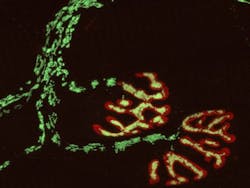
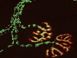
Reactive oxygen species are important intracellular signaling molecules, but their mode of action is complex: In low concentrations, they regulate key aspects of cellular function and behavior, while at high concentrations they can cause "oxidative stress," which damages organelles, membranes, and DNA. To analyze how redox signaling unfolds in single cells and organelles in real time, the optical microscopy technique developed jointly by the teams of LMU professor Martin Kerschensteiner and TUM professor Thomas Misgeld, both investigators of the Munich Cluster for Systems Neurology (SyNergy), allows them to record the oxidation states of individual mitochondria with high spatial and temporal resolution. Kerschensteiner explains the motivation behind the development of the technique: “Redox signals have important physiological functions, but can also cause damage, for example when present in high concentrations around immune cells.”
In earlier studies, Kerschensteiner and Misgeld had obtained evidence that oxidative damage of mitochondria might contribute to the destruction of axons in inflammatory diseases such as multiple sclerosis.
Kerschensteiner and Misgeld used redox-sensitive variants of the green fluorescent protein (GFP) as visualization tools. “By combining these with other biosensors and vital dyes, we were able to establish an approach that permits us to simultaneously monitor redox signals together with mitochondrial calcium currents, as well as changes in the electrical potential and the proton (pH) gradient across the mitochondrial membrane,” says Misgeld.
The researchers have applied the technique to two experimental models, and have arrived at some unexpected insights. On the one hand, they have been able, for the first time, to study redox signal induction in response to neural damage—in this case, spinal cord injury—in the mammalian nervous system. The observations revealed that severance of an axon results in a wave of oxidation of the mitochondria, which begins at the site of damage and is propagated along the fiber. Furthermore, an influx of calcium at the site of axonal resection was shown to be essential for the ensuing functional damage to mitochondria.
Perhaps the most surprising outcome of the new study was that the study's first author, graduate student Michael Breckwoldt, was able to image, also for the first time, spontaneous contractions of mitochondria that are accompanied by a rapid shift in the redox state of the organelle. As Misgeld explains, “This appears to be a fail-safe system that is activated in response to stress and temporarily attenuates mitochondrial activity. Under pathological conditions, the contractions are more prolonged and may become irreversible, and this can ultimately result in irreparable damage to the nerve process.”
Full details of the team's work appear in the journal Nature Medicine; for more information, please visit http://dx.doi.org/10.1038/nm.3520.
-----
Don't miss Strategies in Biophotonics, a conference and exhibition dedicated to development and commercialization of bio-optics and biophotonics technologies!
Follow us on Twitter, 'like' us on Facebook, and join our group on LinkedIn
Subscribe now to BioOptics World magazine; it's free!

