Fluorescence microscopy method enables rapid, whole-brain imaging with single-cell resolution
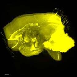
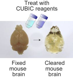
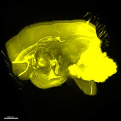
Whole-brain imaging at single-cell resolution normally involves preparing a highly transparent sample that minimizes light scattering and then imaging neurons tagged with fluorescent probes at different slices to produce a 3D representation. However, limitations in current methods prevent comprehensive study of the relationship. Recognizing this, researchers at the RIKEN Quantitative Biology Center (Wako, Japan) have developed a new high-throughput method that they call clear, unobstructed brain imaging cocktails and computational analysis (CUBIC), which offers unprecedented rapid, whole-brain imaging at single-cell resolution and a simple protocol to clear and transparentize the brain sample based on the use of aminoalcohols.
Related: Sugar solution makes tissues see-through, enables images with unprecedented detail
Combined with light-sheet fluorescence microscopy, CUBIC was tested for rapid imaging of a number of mammalian systems, such as mouse and primate, showing its scalability for brains of different size. Additionally, it was used to acquire new spatial-temporal details of gene expression patterns in the hypothalamic circadian rhythm center. And by combining images taken from opposite directions, CUBIC enables whole-brain imaging and direct comparison of brains in different environmental conditions.
CUBIC overcomes a number of obstacles compared with previous methods. One is the clearing and transparency protocol, which involves serially immersing fixed tissues into just two reagents for a relatively short time. Second, CUBIC is compatible with many fluorescent probes because of low quenching, which allows for probes with longer wavelengths and reduces concern for scattering when imaging the whole brain while inviting multicolor imaging. Finally, it is highly reproducible and scalable.
CUBIC provides information on previously unattainable 3D gene expression profiles and neural networks at the systems level. Because of its rapid and high-throughput imaging, CUBIC offers opportunity to analyze localized effects of genomic editing. It also is expected to identify neural connections at the whole-brain level.
In the future, explains center director Hiroki Ueda, MD, Ph.D., the research team would like to apply CUBIC to whole-body imaging at single-cell resolution.
Full details of the work appear in the journal Cell; for more information, please visit http://dx.doi.org/10.1016/j.cell.2014.03.042.
-----
Follow us on Twitter, 'like' us on Facebook, and join our group on LinkedIn
Subscribe now to BioOptics World magazine; it's free!


