Light microscopy trailblazers win Nobel Prize in Chemistry 2014
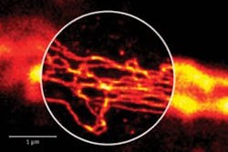
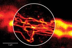
Three light microscopy pioneers—Eric Betzig, Stefan W. Hell, and William E. Moerner—have been awarded the Nobel Prize in Chemistry 2014 for two separate achievements in breaking the optical diffraction limit, which enables better resolution (>0.2 µm) than half the wavelength of light. With the help of fluorescent molecules, the prizewinners were each able to circumvent this limitation.
Related: Nobel Prize honors super-resolution optical microscopy
Hell developed the stimulated emission depletion (STED) microscopy method in 2000, which uses two laser beams—one stimulates fluorescent molecules to glow while another cancels out all fluorescence, except for that in a nanometer-sized volume. Scanning over the sample, nanometer for nanometer, yields an image with a resolution better than the diffraction limit.
Betzig and Moerner, working separately, laid the foundation for the second method, single-molecule microscopy. The method relies upon the possibility to turn the fluorescence of individual molecules on and off. Scientists can image the same area multiple times, letting just a few interspersed molecules glow each time. Superimposing these images yields a dense super-resolution image. In 2006, Betzig utilized this method for the first time.
Both methods are part of what has become known as nanoscopy, which allows scientists to visualize the pathways of individual molecules inside living cells. They can see how molecules create synapses between nerve cells in the brain; track proteins involved in Parkinson's, Alzheimer's, and Huntington's diseases as they aggregate; and follow individual proteins in fertilized eggs as these divide into embryos.
For more information, please visit www.nobelprize.org/nobel_prizes/chemistry/laureates/2014.
-----
Follow us on Twitter, 'like' us on Facebook, connect with us on Google+, and join our group on LinkedIn
Subscribe now to BioOptics World magazine; it's free!

