Super-res microscope explores living cells in 3D noninvasively
A startup company founded last year at the École Polytechnique Federale de Lausanne (EPFL) Innovation Park (Lausanne, Switzerland), Nanolive SA, has developed a super-resolution microscope that enables exploration of a living cell in 3D without damaging it.
Related: Light absorption indicates oxidative stress in cells
While scientists may still obtain a finer resolution using an electron microscope, this approach cannot be used to examine cells that are alive. For a long time, it was believed to be impossible to look inside a living cell using light microscopes due to their physical limitations. This year's Nobel Prize for Chemistry was awarded to Stefan Hell, Eric Betzig, and William Moerner, who did not believe these presumed limitations and made revolutionary discoveries in the field of fluorescence microscopy.
While their research was focused on the chemistry of single molecules and their pathways inside living cells, Nanolive focuses on the physical structure of the living cell itself.
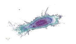
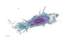
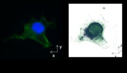
The 3D Cell Explorer super-resolution microscope peers into living cells without sample preparation, enabling noninvasive observation of nuclei and organelles down to 70 nm. Similar to a magnetic resonance imaging (MRI)/computed tomography (CT) scan, the microscope takes a complete tomographic image of the refractive index within living cells, allowing researchers to see and precisely measure the impact of stimuli and drugs on cells.
For more information, please visit http://nanolive.ch/product.
-----
Follow us on Twitter, 'like' us on Facebook, connect with us on Google+, and join our group on LinkedIn
Subscribe now to BioOptics World magazine; it's free!


