Pen-sized device employs spectroscopy methods to boost skin cancer detection
Researchers in the Cockrell School of Engineering at the University of Texas at Austin (UT Austin) have designed a probe that employs a combination of spectroscopy methods to reduce unnecessary biopsies by offering a fast, noninvasive, and lower-cost solution to detect melanoma and other skin cancer lesions.
Related: Verisante launches rapid Raman spectroscopy system for cancer research, other markets
James Tunnell, an associate professor in the Department of Biomedical Engineering, led the researchers in developing the probe, which they have begun testing in clinical trials. They are also partnering with funding agencies and startup companies to help bring the device to dermatologists.
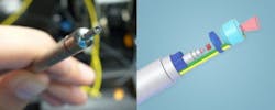
Tunnell and his colleagues combined Raman spectroscopy, diffuse reflectance spectroscopy, and laser-induced fluorescence spectroscopy to create a more complete picture of a skin lesion. By revealing information invisible to the human eye, the probe could offer a faster, better screening tool for cancer and eliminate many biopsies. The probe itself is about the size of a pen, and the spectroscopic and computer equipment that supports it fits onto a portable utility cart that can be wheeled between rooms. Each reading takes about 4.5 s to perform.

As normal skin becomes cancerous, cell nuclei enlarge, the top layers of skin can thicken, and the skin cells can increase their consumption of oxygen and become disorganized, Tunnell explains. The changes alter the way light interacts with the tissue.
To detect all these changes requires multiple spectroscopic techniques. For example, diffuse reflectance spectroscopy is sensitive to absorption by proteins, such as hemoglobin. Raman spectroscopy is sensitive to vibrational modes of chemical bonds, such as those found in connective tissues, lipids, and cell nuclei.
DermDx, a firm seeking to commercialize the research described in this paper, has a technology licensing agreement with UT Austin.
Full details of the research team's work appear in the Review of Scientific Instruments; for more information, please visit http://dx.doi.org/10.1063/1.4890199.
-----
Don't miss Strategies in Biophotonics, a conference and exhibition dedicated to development and commercialization of bio-optics and biophotonics technologies!
Follow us on Twitter, 'like' us on Facebook, and join our group on LinkedIn
Subscribe now to BioOptics World magazine; it's free!