Fluorescence microscopy technique could lead to new drug development against osteoporosis

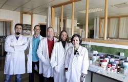
Scientists from the University of Granada (UGR)'s Department of Physio-Chemistry in Spain have turned to a fluorescence microscopy technique that allows specialists to measure the concentration of phosphate ions inside living cells. The technique can evaluate the bioavailability of drugs used in certain illnesses, including osteoporosis—a progressive bone disease that affects bone density, particularly in women over 65.
Related: FTIR spectroscopy helps predict bone density, fracture risk
Related: Speeding the path to clinical trials: optical imaging in drug development
Currently, there are only invasive treatments to calculate phosphate concentration within osteoblasts, which are the precursors to bone cells. To do this, radioactive phosphorus is used, which has serious drawbacks. But the fluorescence microscopy technique that the researchers used—fluorescence lifetime imaging microscopy (FLIM)—can make these calculations noninvasively and in real time. FLIM is based on using a substance that emits fluorescence, generated via prior agitation using a pulsed laser. The time the fluorescence lasts is a signal of the phosphate concentration within the cellular cytoplasm.
The work has been patented via the University of Granada’s Research Results Transference Office (OTRI). Currently, the researchers are looking for pharmaceutical companies that are currently working on the development of drugs to measure the bioavailability of phosphate.
Full details of the researchers' work appear in the Journal of Physical Chemistry B; for more information, please visit http://dx.doi.org/10.1021/jp405041c.
-----
Follow us on Twitter, 'like' us on Facebook, and join our group on LinkedIn
Subscribe now to BioOptics World magazine; it's free!