GENOMICS/MICROSCOPY: Microscope/fluorophore combination enables gene splicing observation
Understanding the function of intracellular micro-machines called spliceosomes is important for basic biology and pathology—and may lead to therapies that fix the splicing process in cases where it is not working properly, says Aaron Hoskins, a postdoctoral fellow at Brandeis University, where researchers have developed a way to use lasers to study, in unprecedented detail, the splicing of pre-messenger RNA molecules.
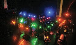
A paper describing the approach is based on a five-year collaboration of three research laboratories. In addition to Hoskins, authors include Jeff Gelles, whose lab developed the multi-laser total internal reflection fluorescence (TIRF) imaging system; Larry Friedman, a key contributor in building the microscope; and researchers at Columbia University (who made specialized probes to tag the molecules) and the University of Massachusetts Medical School.1
Approximately 100 custom and commercial off-the-shelf components comprise the microscope; Friedman has spent more than five years developing specialized light microscopes to study single protein molecules, while Hoskins has been developing the methodology to study these proteins in the complex environments necessary for spliceosome function. “If we have one component of the spliceosome that has a green dye on it and one that has a red dye on it, then we see a green spot and a red spot coming together on the RNA, we know that we are studying part of that assembly process,” says Gelles. “By looking at individual molecules one at a time, we can actually follow the stages of the assembly process. We can determine whether it happens in the same order on each molecule, or if some spliceosomes assemble differently than others.”
According to Hoskins, the most challenging aspect of the project was determining two completely orthogonal methods for attaching fluorophores to endogenous spliceosomes in whole cell extract. “Since these experiments are quantitative, we needed to find methods that give a very high degree of fluorophore incorporation and specificity,” he notes. “In other words, 10 percent labeling would not cut it! The novel part, for me, is that for decades spliceosome kinetics have been ‘off-limits’ to enzymologists due to the complexity of the system. However, by developing the correct analytical tools, the spliceosome can be studied in detail usually reserved for enzymes orders of magnitude smaller.”
The technology the researchers developed is generally applicable to study a wide range of biological problems, says Gelles. “It really enables us to find things out that were very difficult to study using previously existing approaches.” The Gelles lab is now researching the process of RNA transcription, and processes by which cells change their shape and move.
1. A.A. Hoskins et al., Science 331 (6022): 1289–1295 (2011).
More BioOptics World Current Issue Articles
More BioOptics World Archives Issue Articles

