Green fluorescent protein boosts light in first 'bio laser'
Researchers at Harvard Medical School and Massachusetts General Hospital (both in Boston, MA) haveâfor the first timeâcreated laser light using a single living human cell and green fluorescent protein (GFP), the substance that makes jellyfish bioluminescent and is used in cell labeling.
Reported today in Nature Photonics1, Seok-Hyun Yun, an optical physicist at Harvard Medical School and Massachusetts General Hospital in Boston, created the 'bio laser' with his colleague, Malte Gather.
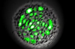
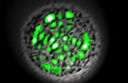
Instead of using doped crystals, semiconductors or gases as the laser's gain medium, Yun and Gather used enhanced GFP. They engineered human embryonic kidney (HEK) cells to produce the GFP, and then placed a single cell between two mirrors to make an optical cavity 20 µm across. When they fed the cell pulses of blue light, it emitted a directional laser beam visible with the naked eye without harming the cell.
However, the researchers' discovery requires further work, including developing the laser so that it works inside an actual living organism. Yun thinks that integrating a nanoscale optical cavity into the laser cell itself could achieve the goal. Technologies to make such cavities are emerging and once they are available, they could be used to create a cell that could "self-lase" from inside tissue, he says.
Michael Berns, a biomedical engineer at the University of California, Irvine, says that the technique might more feasibly be used to study individual cells than for medical applications. He points out that external light is needed to stimulate the laser action, which would be difficult in the body, potentially limiting the technique to thin-tissue systems or cells in culture or suspension.
1. M.C. Gather and S.H. Yun, Nature Photon., doi:10.1038/NPHOTON.2011.99 (2011).
-----
Posted by Lee Mather
Follow us on Twitter, 'like' us on Facebook, and join our group on LinkedIn
Follow OptoIQ on your iPhone; download the free app here.
Subscribe now to BioOptics World magazine; it's free!

