Chemical reagent, fluorescence microscopy team to produce mouse brain tissue details in 3-D
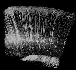
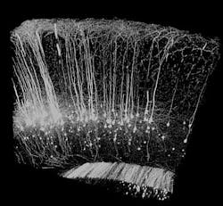
When coupled with fluorescence microscopy, a new aqueous reagent developed by researchers at the RIKEN Brain Science Institute (BSI; Wako City, Japan) can make biological tissue transparent. The team's experiments using fluorescence microscopy on samples treated with the reagent have produced vivid 3-D images of neurons and blood vessels deep inside a mouse brain. Highly effective and cheap to produce, the reagent offers an ideal means for analyzing the complex organs and networks that sustain living systems.
Published in Nature Neuroscience, the new reagent, referred to as Scale and developed by Atsushi Miyawaki and his RIKEN BSI team, renders biological tissue transparent without altering the overall shape or proportions of the sample. Scale also avoids decreasing the intensity of signals emitted by genetically encoded fluorescent proteins in the tissue, which are used as markers to label specific cell types. The combination enables researchers to visualize fluorescently labeled brain samples at a depth of several millimeters and reconstruct neural networks at sub-cellular resolution. Miyawaki and his team have already used Scale to study neurons in the mouse brain at an unprecedented depth and level of resolution, shedding light onto the intricate networks of the cerebral cortex, hippocampus and white matter. Initial experiments exploit Scale's unique properties to visualize the axons connecting left and right hemispheres and blood vessels in the postnatal hippocampus in greater detail than ever possible.
Miyawaki explains that current experiments are focused on the mouse brain, but applications are neither limited to mice, nor to the brain. The team envisions using Scale on other organs such as the heart, muscles, and kidneys, and on tissues from primate and human biopsy samples.
Looking ahead, Miyawaki's team is currently investigating another milder candidate reagent, which would allow them to study live tissue in the same way and at somewhat lower levels of transparency.
-----
Follow us on Twitter, 'like' us on Facebook, and join our group on LinkedIn
Follow OptoIQ on your iPhone; download the free app here.
Subscribe now to BioOptics World magazine; it's free!


