Quantitative imaging, optical microscopy pair to study living bacteria
Researchers from the Université de Paris-Sud (Sceaux, France) and CNRS Montpellier (Montpellier, France) used quantitative imaging (QI) to characterize living bacteria without any immobilization.
Related: Fluorescence imaging sees pigments inside live bacteria cells
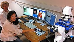
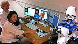
Dr. Christian Marlière of the Institute of Molecular Sciences (ISMO) located at the Université Paris Sud, who led the research team, studied the dynamics and properties of living bacteria in purely controlled physiological conditions at the nanometer scale using atomic force microscopy (AFM) combined with optical microscopy methods having high spatial and temporal resolution, including fluorescence confocal microscopy, photo-activated localization microscopy (PALM), and total internal reflection fluorescence (TIRF) microscopy. Such dynamics and properties included motility or adhesion processes of bacteria on solid substrates, biofilms formation, the influence of light on moving properties of cyanobacteria, and the effects of antibiotics on bacterial membranes and biofilm, among others. Of particular importance was the correlation between the physical, chemical, and biological processes involved at the sub-micrometer scale around bacteria and their biofilms, plus the resulting variation of electrical signals as measured with common macroscopic methods such as electrical conductivity, impedance, or spontaneous potential measurements currently used in geophysics.
Marlière says that using AFM (JPK Instruments' NanoWizard 3 system with a QI mode) in addition to optical microscopy methods enabled them to make measurements of local mechanical, electric, and electro-chemical properties at very well controlled spots on or over the bacteria. Also, he explains, they were able to image living bacteria by AFM in their physiological liquid environment without any external immobilization step. What's more, AFM allowed them to view the native gliding movements of some bacteria (such as cyanobacteria) and obtain important information about the gliding mechanism, he adds.
The lead author of the work was Samia Dhahri, one of Marlière's PhD students. For more information on the work, which appears in the journal PLoS ONE, please visit www.plosone.org/article/info%3Adoi%2F10.1371%2Fjournal.pone.0061663.
-----
Follow us on Twitter, 'like' us on Facebook, and join our group on LinkedIn
Subscribe now to BioOptics World magazine; it's free!

