Fluorescent probes light up live synapses, show how memories form
Scientists at the University of Southern California (USC; Los Angeles, CA) engineered microscopic probes that light up synapses in a living neuron in real time by attaching fluorescent markers onto synaptic proteins, all without affecting the neuronâs ability to function. The fluorescent markers allow scientists to see live excitatory and inhibitory synapses for the first time and, importantly, how they change as new memories are formed.
Related: Optogenetics shows that memories live in specific brain cells
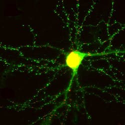
The synapses appear as bright spots along dendrites (the branches of a neuron that transmit electrochemical signals). As the brain processes new information, those bright spots change, visually indicating how synaptic structures in the brain have been altered by the new data.
âWhen you make a memory or learn something, thereâs a physical change in the brain. It turns out that the thing that gets changed is the distribution of synaptic connections,â says Don Arnold, associate professor of molecular and computational biology at the USC Dornsife College of Letters, Arts and Sciences, who co-led the work.
The probes behave like antibodies, but they bind more tightly and are optimized to work inside the cellâsomething that ordinary antibodies canât do. To make these probes, the team used a technique known as âmRNA display,â which was developed by Richard Roberts and Nobel laureate Jack Szostak.
âUsing mRNA display, we can search through more than a trillion different potential proteins simultaneously to find the one protein that binds the target the best,â said Roberts, professor of chemistry and chemical engineering with joint appointments at USC Dornsife and the USC Viterbi School of Engineering, who led the team along with Arnold.

Arnold and Robertsâ probesâdubbed âFingRsââare attached to green fluorescent protein (GFP), a protein isolated from jellyfish that fluoresces bright green when exposed to blue light. Because FingRs are proteins, the genes encoding them can be put into brain cells in living animals, causing the cells themselves to manufacture the probes.
The design of FingRs also includes a regulation system that cuts off the amount of FingR-GFP that is generated after 100 percent of the target protein is labeled, effectively eliminating background fluorescenceâgenerating a sharper, clearer picture.
These probes can be put in the brains of living mice and then imaged through cranial windows using two-photon microscopy.
The new research could offer crucial insight for scientists responding to President Barack Obamaâs Brain Research Through Advancing Innovative Neurotechnologies (BRAIN) Initiative, which was announced in April 2013. Modeled after the Human Genome Project, the objective of the $100 million initiative is to fast-track research that maps out exactly how the brain works and âbetter understand how we think, learn and remember,â according to the BRAIN Initiative website.
The research appears in the journal Neuron; for more information, please visit http://www.cell.com/neuron/abstract/S0896-6273%2813%2900319-X.
-----
Follow us on Twitter, 'like' us on Facebook, and join our group on LinkedIn
Subscribe now to BioOptics World magazine; it's free!