Multispectral fluorescence camera detects tumors
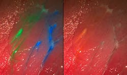
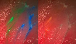
Researchers at the Fraunhofer Project Group for Automation in Medicine and Biotechnology (PAMB), a project that belongs to the Fraunhofer Institute for Manufacturing Engineering and Automation (IPA; Stuttgart, Germany), have developed a multispectral fluorescence camera system that could someday integrate into medical imaging systems such as surgical microscopes and endoscopes. Their camera is the first of its kind able to display several fluorescent dyes and the reflectance image simultaneously in real time, enabling healthy arteries and delicate nerves to be colored with dye and then be detected.
Related: Multispectral system enables brain development discovery
âThe visibility of the dye to the camera depends in large part on the selection of the correct set of fluorescence filters. The filter separates the incident excitation wavelengths from the fluorescing wavelengths so that the diseased tissue is also set apart from its surroundings, even at very low light intensities,â says Nikolas Dimitriadis, head of the Biomedical Optics Group at PAMB.
The researchers and their colleagues require only one camera and one set of filters for their photographs, which can present up to four dyes at the same time. Software developed in-house analyzes and processes the images in seconds and presents it continuously on a monitor during surgery. The information from the fluorescent image is superposed on the normal color image.
âThe operator receives significantly more accurate information. Millimeter-sized tumor remnants or metastases that a surgeon would otherwise possibly overlook are recognizable in detail on the monitor. Patients operated under fluorescent light have improved chances of survival,â says Dimitriadis.
In order to make the multispectral fluorescence camera system as adaptable as possible, it can be converted to other combinations of dyes. âOne preparation that is already available to make tumors visible is 5-amino levulinic acid (5-ALA). Physicians employ this especially for glioblastomasâone of the most frequent malignant brain tumors in adults,â explains Dimitriadis. 5-ALA leads to an accumulation of a red dye in the tumor and can likewise be detected with the camera.
The scientists expect that their camera should pass testing for use with humans as soon as next year. The first clinical tests with patients suffering from glioblastomas are planned for 2014.
They will debut their camera prototype at the Medica Trade Fair, to take place November 20-23, 2013, in Düsseldorf, Germany, in the joint Fraunhofer booth (Halle 10, Booth F05).
-----
Follow us on Twitter, 'like' us on Facebook, and join our group on LinkedIn
Subscribe now to BioOptics World magazine; it's free!

