Three-photon microscopy boosts biological imaging deep in the brain
Researchers at Cornell University (Ithaca, NY) have broken the fundamental depth limit of standard two-photon microscopy, demonstrating high-resolution, 3D imaging of the subcortical region of a live, intact mouse brain. The ability to image that deep and with that much detail could lead to deeper understanding of how the brain works, including finding cures for depression, Alzheimer's, and Parkinson's diseases.
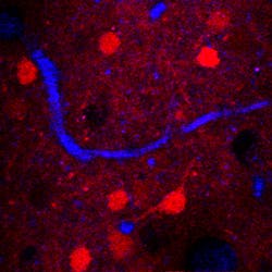
The research teamâled by Chris Xu, associate professor of applied and engineering physicsâused a mouse model to prove the principle of three-photon microscopy operating at a 1,700 nm wavelength. Using a new laser specifically created for three-photon excitation allowed the researchers to perform high-resolution imaging of neurons at unprecedented depths within a mouse brain. Three-photon fluorescence, combined with the longer excitation wavelength of the laser pulse, enabled the researchers to overcome obstacles such as tissue scattering and absorption, which prohibit high-resolution imaging deep within biological tissues.

In the mouse model, the researchers used dyes and transgenic mice to test their multiphoton microscope on different fluorescent signals and prove their concept. If three-photon microscopy can be used to map the entire mouse brain, it could ultimately help shed light on the functions of human brains and pave the way to breakthroughs in neuroscience and other clinically relevant areas, Xu says.
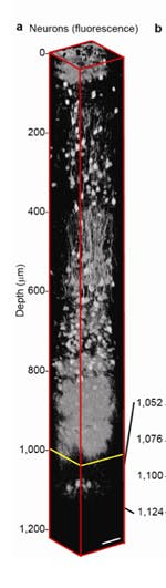
"Brain mapping could be the so-called grand challenge within the next decade," Xu says. "With MRI, we can see the whole brain but not with the resolution we have demonstrated. The optical resolution is about 100 to 1,000 times higher and allows us to clearly visualize individual neurons."
Full details of the team's work appear in Nature Photonics; for more information, please visit http://www.nature.com/nphoton/journal/vaop/ncurrent/full/nphoton.2012.336.html.
-----
Follow us on Twitter, 'like' us on Facebook, and join our group on LinkedIn
Subscribe now to BioOptics World magazine; it's free!