Single-molecule imaging could get boost from nanoscale technology to study diseases

July 22, 2016
A low-cost imaging platform enables single-molecule imaging, and could transform study of molecules and cells at nanoscale levels.

A team of researchers at the University of Missouri (Columbia, MO) has developed a relatively inexpensive imaging platform that enables single-molecule imaging and, therefore, could transform how scientists study molecules and cells at nanoscale levels.

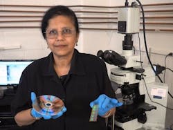
Shubra Gangopadhyay, an electrical and computer engineer at the University of Missouri College of Engineering, recently published studies outlining a new, relatively inexpensive imaging platform that enables single-molecule imaging.

Shubra Gangopadhyay, an electrical and computer engineer at the University of Missouri College of Engineering who led the work, explains that scientists usually have to use very expensive microscopes to image at the sub-microscopic level. So the techniques she and her team have established, she says, help to produce enhanced imaging results with ordinary microscopes. What's more, the low cost to produce their platform also means it could be used to detect a wide variety of diseases, particularly in developing countries.

A schematic showing the difference between regular and plasmonic gratings in terms of fluorescence intensity. (Photo courtesy of Shubhra Gangopadhyay/Nanoscale)

The custom platform uses an interaction between light and the surface of the metal grating to generate surface plasmon resonance (SPR), a rapidly developing imaging technique that enables super-resolution imaging down to 65 nm—a resolution normally reserved for electron microscopes. Using HD-DVD and Blu-ray discs as starting templates, a repeating grating pattern is transferred onto the microscope slides where the specimen will be placed. Since the patterns originate from a widely used technology, the manufacturing process remains relatively inexpensive. In previous studies, Gangopadhyay says, she and her team used plasmonic gratings to detect cortisol and tuberculosis.

Full details of the work appear in the journal Nanoscale; for more information, please visit http://dx.doi.org/10.1039/C5NR09165A.

About the Author

BioOptics World Editors

We edited the content of this article, which was contributed by outside sources, to fit our style and substance requirements. (Editor’s Note: BioOptics World has folded as a brand and is now part of Laser Focus World, effective in 2022.)

Sponsored Recommendations

Looking beyond the incremental: Reimagining optical devices

Dec. 7, 2023
Rob Devlin, co-founder and CEO of Metalenz, shares the significant people that forged his path to developing one of the most promising optics companies in the world today.

Monolithic integration of functional structures into micro-optical elements

Dec. 6, 2023
A polymer-only ultraviolet imprint process potentially saves costs, simplifies the process, and increases the reliability of the optical element.

Manufacturing thin films with tailor-made electronic properties

Dec. 5, 2023
Unlock the future of optoelectronics as researchers at Leibniz IPHT in Jena, Germany unveil an innovative technique for precision deposition of thin organic semiconductor films...

Quantitative Microscopy with Deep Learning

Dec. 5, 2023
Explore the untapped potential of deep learning in video microscopy with our cutting-edge software, DeepTrack 2.2. Overcoming the steep learning curve, this innovative application...

Voice your opinion!

To join the conversation, and become an exclusive member of Laser Focus World, create an account today!