Fluorescence approach activates glutamate receptors to better understand brain pathology

The α-amino-3-hydroxy-5-methyl-4-isoxazolepropionic acid (AMPA)-type glutamate receptor ensures that neurotransmission proceeds at a breakneck pace from cell to cell in the brain. Seeking to understand its activation mechanisms, a team of researchers at the Leibniz Institute for Molecular Pharmacology (FMP; Berlin, Germany) used fluorescence resonance energy transfer (FRET; a fluorescence imaging approach) to watch these activated receptors at work. By doing so, the researchers detected movements of unexpected parts of the receptor optically and electrically at the same time, which could lead to new ways to see into the brain—including in pathological situations.
AMPA-type glutamate receptors (AMPARs) in the brain ensure that neurotransmitter messages are transmitted across synapses with extreme speed. This lightning-fast synaptic transmission is essential for everything we do, think, and remember, so AMPARs are the most common excitatory receptors in the brain and essential for our survival.
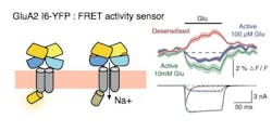
Although the binding of neurotransmitters to AMPARs is quite well understood, its activation mechanisms have not been fully unraveled. So, the research team—led by Dr. Andrew Plested and PhD student Ljudmila Katchan—used FRET to make the activation of AMPARs optically visible. They found that neurotransmitters drive conformational changes in two intracellular regions of the receptor, as well as at extracellular location where the glutamate binds to the receptor. Even more, the researchers were able to watch the receptor activity in real time. The motivation for this research is that given a receptor that has big enough changes in its light output, they could also watch synapses.
Research group leader Plested explained that his team's purpose with their work was to simultaneously measure activation optically and electronically. Collaborators from Anders Kristensen's team at the University of Copenhagen (Denmark) first saw definite optical signals with fluorescence-lifetime imaging microscopy (FLIM), but this method is too slow to see activation. "We have provided the proof of principle, opening the door to use this technique for research into the functional properties of other signaling molecules in the brain," Plested says.
The researchers now want to investigate other molecules essential for neurotransmission, beginning with cell cultures and later in the brain. Even if all (billions) synapses in the brain cannot be visualized at once with the new method, following just a handful of them could give new insights into thinking, Plested explains. Moreover, he says, these approaches could one day help to gain a better understanding about neurodegenerative diseases, including the activity or function of the lost synapses and the ones that remain.
Full details of the work appear in the Proceedings of the National Academy of Sciences; for more information, please visit http://dx.doi.org/10.1073/pnas.1601747113.