Combo fluorescence approach can monitor two signaling molecules within a dendritic spine

A team of researchers at the Max Planck Florida Institute for Neuroscience (MPFI; Jupiter, FL) has been working to understand the cellular and molecular mechanisms of learning and memory. They have been combining two fluorescence imaging techniques—fluorescence resonance energy transfer (FRET) and fluorescence lifetime imaging microscopy (FLIM)—to study the activity of proteins in dendritic spines (protrusions that form off of neuronal branches), and make synaptic connections and communicate with other neurons. Dendritic spines are known to emerge, change shape, and even disappear over a lifetime, and these changes are considered the cellular basis for learning and memory. These imaging techniques were a key factor in helping the team elucidate some of the molecular mechanisms behind this type of plasticity.
Related: Fluorescence imaging method visualizes nine structures in one measurement
However, there is an important limitation in using these techniques to understand how multiple types of proteins and molecules interact in living samples. Green fluorescent protein (GFP) is the only FRET donor tag that will work within a FLIM protocol, so if researchers want to study two proteins, they'll investigate "Protein A" in one set of experiments, following an additional set of experiments to examine "Protein B" activity. Following these experiments, the researchers will need to draw conclusions about how these two proteins interact based on these two sets of experiments. This not only increases the amount of time it takes to study multiple proteins, but also makes it more difficult to analyze how they interact in space and time. "For those types of systems, it's crucial to look at as many proteins as possible at the same time to correlate their activities," explains Tal Laviv, Ph.D., a postdoctoral researcher in MPFI scientific director Ryohei Yasuda's laboratory, which led the work.
The research team of Dr. Michael Lin at Stanford University (California), which specializes in building protein-based tools for molecular imaging, reached out to the Yasuda Lab at MPFI after they identified a new set of red fluorescent proteins (RFPs) named CyRFPs. They suggested this new set of RFPs could be used in combination with GFP for simultaneous imaging.
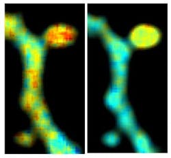
To determine if this could work, the team of scientists tested the ability to visualize dendritic spine structure and function using CyRFP and a GFP-based calcium biosensor. Using this combination, they were able to monitor the structure and function of spines in real time, even in the brains of living animals. Finally, the team tweaked a variant of CyRFP, which now could be used as a fluorescent FRET donor a part of a FRET pair, named monomeric cyan-excitable RFP (mCyRFP1). Scientists in the Yasuda Lab conducted a series of experiments to test the newly proposed FRET pair alongside a GFP biosensor. The technique allowed them to view the activities of two signaling molecules within a single dendritic spine as the spine was undergoing synaptic plasticity.
Laviv explains that the new technique will increase both accuracy and efficiency of FRET-FLIM imaging experiments and could potentially increase our understanding of how learning and memory ultimately alters the structure and function of dendritic spines.
Full details of the work appear in the journal Nature Methods (doi:10.1038/nmeth.4046).