Researchers detail transparent graphene sensor technology for bioimaging, optogenetics
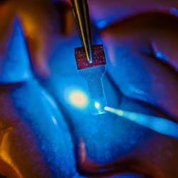
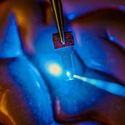
In a new paper published in the journal Nature Protocols, a team of researchers at the University of Wisconsin-Madison (UW-Madison), which had developed a transparent graphene sensor for use in imaging the brain back in 2014, has described in detail how to fabricate and use the see-through, implantable micro-electrode arrays in fluorescence microscopy, optical coherence tomography (OCT), and optogenetics applications, as well as electrophysiology.
Related: Raman spectroscopy-based graphene sensor could detect viruses
Because many research groups asked the UW-Madison research team for the sensor devices described in the 2014 paper, they were unable to keep up with the requests, according to Zhenqiang (Jack) Ma, the Lynn H. Matthias Professor and Vilas Distinguished Achievement Professor in electrical and computer engineering at UW-Madison. The team, he says, made the decision in their latest paper to describe how to do these things so that they can start working on the next generation of the sensors.
Ma and his collaborator Justin Williams, the Vilas Distinguished Achievement Professor in biomedical engineering and neurological surgery at UW-Madison, patented the technology through the Wisconsin Alumni Research Foundation, seeing its potential for advancements in research. "That little step has already resulted in an explosion of research in this field," Williams says. "We didn't want to keep this technology in our lab. We wanted to share it and expand the boundaries of its applications."
Now, not only are the UW-Madison researchers looking at ways to improve and build upon the technology, they also are seeking to expand its applications from neuroscience into areas such as research of stroke, epilepsy, Parkinson's disease, cardiac conditions, and many others.
To view the full details of the work in the Nature Protocols paper, please visit http://dx.doi.org/10.1038/nprot.2016.127.

