Optoacoustic imaging method can observe large neural circuits in 3D and real time
Most brain functions are not well understood through inspection of single neurons, so neuroscientists need the ability to monitor the activity of millions of neurons, both individually and collectively. However, such observations into the living brain are limited by the penetration depth of optical microscopy techniques.
Related: Optogenetics helps untangle ambiguity in neural circuits
So, a team of researchers from Helmholtz Zentrum München and the Technical University of Munich (both in Munich, Germany) has now found a way to address this challenge. The new method is based on optoacoustics (also known as photoacoustics), which allows noninvasive interrogation of living tissues at centimeter-scale depths by using short laser pulses that cause short-term expansion of the tissue to yield tiny ultrasound vibrations. The vibrations are then registered with specially designed detectors, processed, and converted into three-dimensional (3D) images of the interrogated tissue.
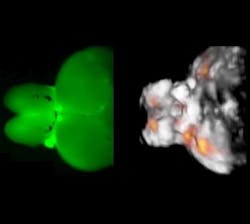
"We discovered that optoacoustics can be made sensitive to the differences in calcium ion concentrations resulting from neural activity and devised a rapid functional optoacoustic neuro-tomography (FONT) system that can simultaneously record signals from a very large number of neurons," says Dr. Xosé Luis Deán-Ben, first author of the paper describing the work. Experiments performed by the scientists in brains of adult zebrafish (Danio rerio) expressing genetically encoded calcium indicator GCaMP5G demonstrated the fundamental ability to directly track neural dynamics using optoacoustics while overcoming the longstanding penetration barrier of optical imaging in opaque brains. The technique was also able to trace neural activity during unrestrained motion of the animals.
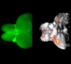
"So far, we demonstrated real-time analysis on whole brains of adult animals with roughly 2 × 3 × 4 mm dimensions (approximately 24 mm3)," says Prof. Dr. Daniel Razansky, a group leader at the Institute of Biological and Molecular Imaging (IBMI) at Helmholtz Zentrum München and Professor of Molecular Imaging Engineering at the Technical University of Munich. State-of-the-art optical microscopy methods are currently limited to volumes well below a cubic millimeter when it comes to imaging of fast neural activity, according to the researchers. In addition, their FONT method is already capable of visualizing volumes of more than 1000 mm3 with temporal resolution of 10 ms.
Large-scale observation of neural activity is the key to understanding how the brain works, both under normal and diseased conditions. "This new imaging tool is expected not only to significantly promote our knowledge on brain function and its pathophysiology, but also accelerate development of novel therapies targeting neurological and neuropsychiatric disorders," Razansky concludes.
Full details of the work appear in the journal Light: Science & Applications; for more information, please visit http://dx.doi.org/10.1038/lsa.2016.201.