Mirror approach boosts super-resolution microscopy for cell studies
By growing cells on tiny mirrors and imaging them using super-resolution microscopy, an international team of researchers from Peking University (Beijing, China), the Georgia Institute of Technology (Georgia Tech; Atlanta, GA), and the University of Technology Sydney (UTS; Australia) was able to see the structures of three-dimensional cells with comparable resolution in each dimension.
Related: Live-cell imaging enables super resolution in space and time
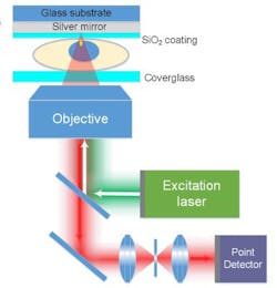
Their technique (which also works with other technologies) uses the unique properties of light to create interference patterns as light waves pass through a cell on the way to the mirror and then back through the cell after being reflected. The interference patterns provide, at a single plane within the cell, significantly improved resolution in the z-axis. This improved view could help differentiate between structures that appear close together with existing microscope technology, but are actually relatively far apart within the cells.
"A single cell is about 10 µm; inside that is a nuclear core about 5 µm, and inside that are tiny holes, called the 'nuclear pore complex,' that as a gate regulates the messenger biomolecules, but measure between one-fiftieth and one-twentieth of a micrometer," says Dayong Jin, a professor at UTS and one of the paper's co-authors. With the technique, he explains, they were able to see the details of those tiny holes.
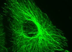
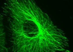
Peng Xi, a professor at Peking University and another of the paper's co-authors, adds that being able to see these tiny structures may provide new information about the behavior of cells, how they communicate, and how diseases arise in them. The new system, he notes, allows scientists to see the ring structure of the nuclear pore complex and the tubular structure of the human respiratory syncytial virus (hRSV).
While changing the optical system was relatively simple, growing cells on the custom-made mirrors required adapting existing biological techniques, says Phil Santangelo, another co-author and a professor at Georgia Tech and Emory University. Techniques for growing the cells on the mirrors were largely developed by Eric Alonas, a Georgia Tech graduate student, and Hao Xie, a student in the PhD program of Peking University and Georgia Tech.
The new technique, known as mirror-enhanced, axial narrowing, super-resolution (MEANS) microscopy, begins with growing cells to be studied on a tiny mirrors custom-made by a manufacturer in China. A glass cover slide is placed over the cells, and the mirror placed into a confocal or wide-field microscope in the place of a usual clear slide.
The technique improves axial resolution six-fold and lateral resolution two-fold for stimulated emission depletion (STED) nanoscopy. The ability to increase the resolution and decrease the thickness of an axial section without increasing laser power is of great importance for imaging biological specimens, which cannot tolerate high laser power, the researchers note.
Santangelo believes the technique could find broad applications for scientists using fluorescence microscopy to examine cells and subcellular structures. Further research could lead to improvements such as the ability to make the mirror's surface movable, allowing more control over how the cells can be imaged.
Full details of the work appear in the journal Light: Science & Applications; for more information, please visit http://dx.doi.org/10.1038/lsa.2016.134.


