Enhanced fluorescence microscopy method could be accessible for most cell biology labs

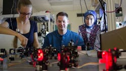
A team of researchers at the University of New Mexico (UNM; Albuquerque, NM), led by associate professor Keith Lidke and working with collaborators at Sandia National Laboratories (also in Albuquerque), has improved upon an existing method of fluorescence microscopy to allow a better view of cellular interactions.
Related: Hyperspectral microscope can view molecular-level protein-protein interactions in living cells
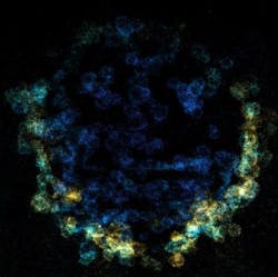
According to Lidke, who works in UNM's Department of Physics & Astronomy, traditional fluorescence microscopy methods can only provide researchers a very limited view of the cells they're looking at, and exposes the sample to an abundance of light that degrades the image quality and leads to cell damage through phototoxicity. "What light-sheet microscopy does is allow us create a sheet of light that is matched exactly to the focal plane that we're imaging," Lidke explains. "We reduce light exposure and we reduce background noise in the system, so in living cells that allows us to see fluorescent proteins with enough signal to look at the dynamics of those proteins."
The research team's method, called single-objective light-sheet microscopy, overcomes these prominent issues and allows light-sheet microscopy to be performed using common microscopes found in most cell biology laboratories.
While Lidke's method is still in its early stages, he has already received a lot of interest from researchers at UNM and across the country because of the unique view the equipment can provide. According to Lidke, cells function through signaling pathways, which are a series of protein-protein interactions—but exactly how those interactions work isn't clear because of a lack of technology available to see those events happen in living cells. The method has the ability to help answer questions about how cells communicate and work internally, making it possible for researchers to develop medicine or therapies that utilize these interactions.
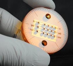
The new method is made possible through two different components—a specialized optics attachment that creates the light sheet and a highly engineered microfluidic chip that holds the sample. Lidke's group is responsible for creating the optics component, which was developed as an attachment to most epifluorescent microscopes as a way to make the technique usable for a large audience. Collaborators at Sandia National Labs worked with the group to develop the microfluidic chip, which has an integrated mirror in it that allows them to create the light sheet using a single-objective lens. Together, these two pieces give researchers the opportunity to see cellular interaction on an entirely new level.
Right now, Lidke says he's working with the team at Sandia to develop an improved, next-generation chip that he expects to be made commercially available to researchers.
Full details of the work appear in the journal Biomedical Optics Express; for more information, please visit http://dx.doi.org/10.1364/boe.7.002219.